RH-1
Cat.No.: CSC-C0504
Species: Homo sapiens (Human)
Morphology: epitheloid cells growing adherently in monolayers
Culture Properties: monolayer
- Specification
- Background
- Scientific Data
- Q & A
- Customer Review
Immunology: cytokeratin -, desmin -, endothel -, GFAP -, neurofilament -, vimentin +
Viruses: PCR: EBV -, HBV -, HCV -, HIV -, HTLV-I/II -, SMRV -
RH-1 Cells are a line of human tumor cells that was originally established from a childhood soft tissue tumor, but later found to be a member of the Ewing sarcoma family of tumors (ESFT) upon molecular characterization. They were originally thought to be rhabdomyosarcoma cells but later gene expression profiling and cytogenetic analysis discovered they contained EWSR1-FLI1 transcripts, leading many to accept RH-1 cells as an in vitro model of Ewing sarcoma.
RH-1 Cells typically form adherent cell cultures with epithelial-like characteristics when maintained in vitro and divide quickly in normal growth conditions. RH-1 cells also express transcriptional programs associated with oncogenic EWS-FLI1, which modulates several downstream targets. Because of this transcriptional activity, RH-1 cells have been used to characterize signaling pathways associated with Ewing sarcoma.
RH-1 Cells have been used to study tumor biology, oncogenic transcription and expression, as well as novel targeted therapeutics. RH-1 Cells have been utilized as a system to understand how EWS-FLI1 regulates transcription to induce proliferation and how it can be chemosensitive. RH-1 cells have also been used in screens for novel anti-cancer therapeutics that target pathways associated with ESFT.
The Differentiation Protocol Allows the In-Vitro Generation of Erythroid Cells from a Wide Variety of Human Stem Cells
Plasmodium falciparum's clinically relevant blood stage involves erythrocyte invasion, yet studying host genetic factors is limited by erythrocytes' anuclear nature. Pance et al. overcame this by generating erythroid cells from stem cells using an in vitro differentiation protocol, combining flow cytometry-based hemozoin detection with patient-derived iPSC reprogramming and genome editing.
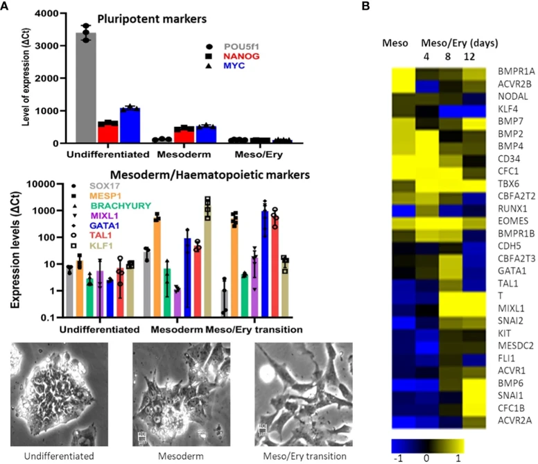
The objective was establishing a protocol to produce sufficient erythroid cells for invasion assays. To minimize cell loss, embryoid body stages and co-cultures were avoided, instead directing cells toward mesoderm through two cytokine steps based on Vallier et al. (2009). First, low Activin A with FGF2 inhibition suppressed neuroectoderm; then BMP4, FGF2, and SB431542 (Activin A inhibition) favored mesoderm over endoderm. IL-3 was added to direct mesoderm toward haematopoiesis. qRT-PCR showed declining pluripotency markers and rising mesoderm markers (Fig. 1A), followed by induction of megakaryocyte-erythroid progenitor transcription factors, coinciding with morphological changes.
Microarray analysis confirmed transcriptome transition from early mesoderm to haematopoiesis (Fig. 1B). Mesoderm specification genes (eomes, BMP2/4, BMPR1B, CFC1) peaked during the Meso-Ery transition, while haematopoietic drivers (T, Tal1, GATA1, MIXL1) peaked later. Based on haematopoietic gene expression, this stage was set at 8-12 days, with cells spontaneously detaching for harvest without trypsinisation.
Ask a Question
Write your own review
- You May Also Need

Description: Organism: Homo sapiens (human)Ethnicity: CaucasianAge/Stage: 36 years of ageGender: maleTissue: sarcomaGrowth Properties: monolayerDescription: in vitro established from a primary skin sarcoma (sole ...

Description: Human cell line derived from epithelioid sarcoma. Derived from a different patient from the patient of HS-ES-1 cell line.

Description: Established from an endometrial stromal sarcoma of the uterus of a 76-year-old Caucasian woman; cells were described as demonstrating a mainly mesenchymal phenotype with signs of epithelial ...

Description: Established from the alveololar rhabdomyosarcoma of the liver of a girl after chemotherapy; cells were described to contain a deletion mutation of p53 and to be resistant to FAS- and TRAIL-induced ...

- Adipose Tissue-Derived Stem Cells
- Human Neurons
- Mouse Probe
- Whole Chromosome Painting Probes
- Hepatic Cells
- Renal Cells
- In Vitro ADME Kits
- Tissue Microarray
- Tissue Blocks
- Tissue Sections
- FFPE Cell Pellet
- Probe
- Centromere Probes
- Telomere Probes
- Satellite Enumeration Probes
- Subtelomere Specific Probes
- Bacterial Probes
- ISH/FISH Probes
- Exosome Isolation Kit
- Human Adult Stem Cells
- Mouse Stem Cells
- iPSCs
- Mouse Embryonic Stem Cells
- iPSC Differentiation Kits
- Mesenchymal Stem Cells
- Immortalized Human Cells
- Immortalized Murine Cells
- Cell Immortalization Kit
- Adipose Cells
- Cardiac Cells
- Dermal Cells
- Epidermal Cells
- Peripheral Blood Mononuclear Cells
- Umbilical Cord Cells
- Monkey Primary Cells
- Mouse Primary Cells
- Breast Tumor Cells
- Colorectal Tumor Cells
- Esophageal Tumor Cells
- Lung Tumor Cells
- Leukemia/Lymphoma/Myeloma Cells
- Ovarian Tumor Cells
- Pancreatic Tumor Cells
- Mouse Tumor Cells