

KYSE-410
Cat.No.: CSC-C0429
Species: Homo sapiens (Human)
Source: Esophagus
Morphology: epitheloid adherent cells growing in monolayers
Culture Properties: monolayer
- Specification
- Background
- Scientific Data
- Q & A
- Customer Review
Immunology: cytokeratin +, cytokeratin-7 +, cytokeratin-8 +, cytokeratin-17 +, cytokeratin-18 +, cytokeratin-
KYSE-410 is a rigorously authenticated human esophageal squamous cell carcinoma (ESCC) cell line established from a poorly differentiated invasive primary tumor resected from the cervical esophagus of a 51-year-old patient prior to treatment. As a founding member of the extensively characterized KYSE series, it offers exceptional model fidelity for ESCC research.
Molecularly, KYSE-410 is distinguished by epigenetic silencing of the p16 (INK4a) tumor suppressor via 5' CpG island hypermethylation while retaining wild-type p15 (INK4b)—a selective inactivation pattern representative of specific ESCC subtypes. The line further harbors pathogenic TP53 and KRAS mutations, MYC amplification, and robust overexpression of hst-1 (heparin-binding growth factor) and cyclin D1, faithfully recapitulating key oncogenic drivers and proliferative signaling circuits.
Cytogenetically, KYSE-410 exhibits a hypotetraploid karyotype with authenticated STR profiling. It demonstrates confirmed tumorigenicity in athymic nude mice, with resultant tumors displaying classical ESCC histology. With comprehensive molecular annotation, standardized culture protocols, and proven utility across in vitro and in vivo platforms, KYSE-410 remains a cornerstone model for investigating ESCC biology, epigenetic therapeutics, and drug development.
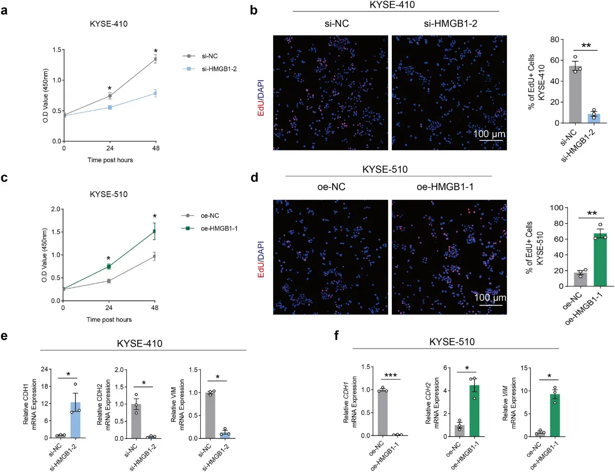
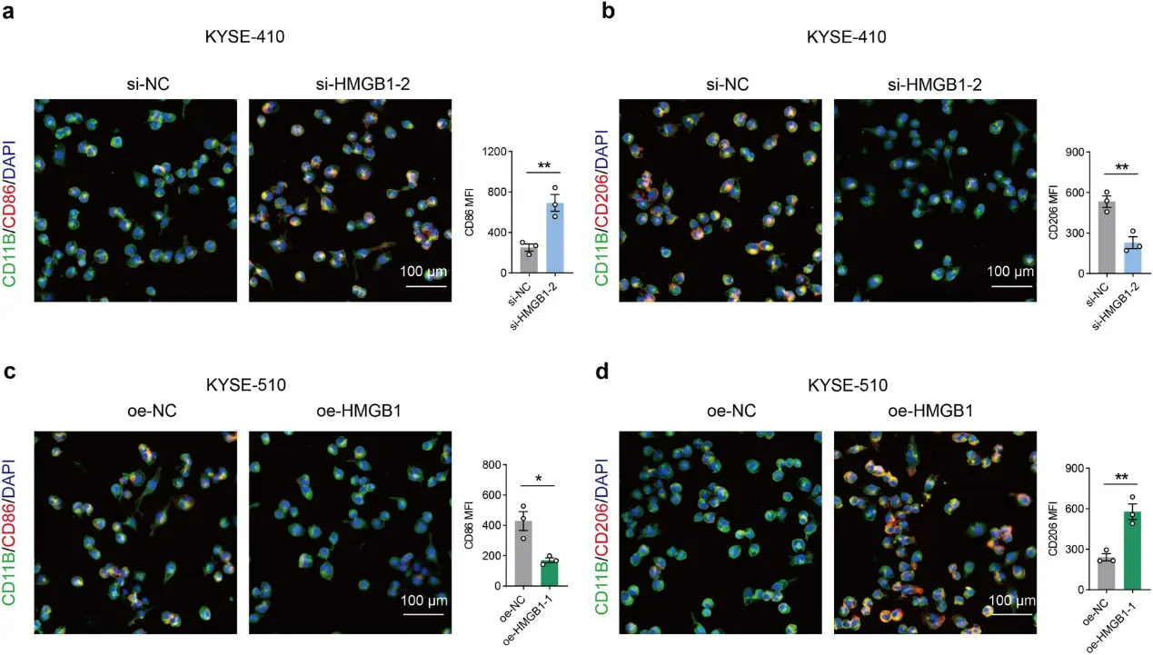
The Mechanism of HMGB1 in the Proliferation and Macrophage Polarization in Esophageal Squamous Cell Carcinoma Cells
Previous studies showed that high mobility group box-1 (HMGB1) facilitates the initiation and progression of esophageal squamous cell carcinoma (ESCC), and the current research investigated the detailed mechanisms implicated.
Quantitative real-time PCR (qRT-PCR) was performed to detect the expressions of HMGB1 in both human esophageal epithelial cells (HEEC) and ESCC cells. After cell transfection, the proliferation of ESCC cells was measured, and the cell metastasis was determined based on the levels of cadherins (CDHs) and Vimentin (VIM). Macrophage polarization was determined by calculating the mean fluorescence intensity (MFI) of CD206 and CD86.
HMGB1 showed a higher expression in ESCC cells, while knockdown of HMGB1 inhibited ESCC cell proliferation, downregulated the levels of CDH2 and VIM and upregulated the level of CDH1. In contrast, overexpressed HMGB1 showed the opposite effects, suggesting the role of HMGB1 in the epithelial–mesenchymal transition (EMT) of ESCC. After the knockout of HMGB1, the MFI of CD86 was increased but that of CD206 was reduced, indicating the polarization towards M1 macrophages. However, the results were reversed when HMGB1 was overexpressed.
Ask a Question
Write your own review
- You May Also Need

Description: Human moderately differentiated squamous cell carcinoma cell line established from esophageal cancer.

Description: Human moderately differentiated squamous cell carcinoma cell line established from esophageal cancer.


Description: Human moderately differentiated squamous cell carcinoma cell line established from esophageal cancer.
Description: Human squamous cell carcinoma from oral cavity via mouse transplantation.

Description: Human squamous cell carcinoma cell line established from recurrent cancer (esophageal cancer).
- Adipose Tissue-Derived Stem Cells
- Human Neurons
- Mouse Probe
- Whole Chromosome Painting Probes
- Hepatic Cells
- Renal Cells
- In Vitro ADME Kits
- Tissue Microarray
- Tissue Blocks
- Tissue Sections
- FFPE Cell Pellet
- Probe
- Centromere Probes
- Telomere Probes
- Satellite Enumeration Probes
- Subtelomere Specific Probes
- Bacterial Probes
- ISH/FISH Probes
- Exosome Isolation Kit
- Human Adult Stem Cells
- Mouse Stem Cells
- iPSCs
- Mouse Embryonic Stem Cells
- iPSC Differentiation Kits
- Mesenchymal Stem Cells
- Immortalized Human Cells
- Immortalized Murine Cells
- Cell Immortalization Kit
- Adipose Cells
- Cardiac Cells
- Dermal Cells
- Epidermal Cells
- Peripheral Blood Mononuclear Cells
- Umbilical Cord Cells
- Monkey Primary Cells
- Mouse Primary Cells
- Breast Tumor Cells
- Colorectal Tumor Cells
- Esophageal Tumor Cells
- Lung Tumor Cells
- Leukemia/Lymphoma/Myeloma Cells
- Ovarian Tumor Cells
- Pancreatic Tumor Cells
- Mouse Tumor Cells