
KB-3-1
Cat.No.: CSC-C6211X
Species: Homo sapiens (Human)
Source: Uterus; Cervix
Morphology: epithelial-like, adherent cell line growing in monolayers
Culture Properties: monolayer
- Specification
- Background
- Scientific Data
- Q & A
- Customer Review
Immunology: cytokeratin +, cytokeratin-7 -, cytokeratin-8 +, cytokeratin-17 +, cytokeratin-18 +, desmin -, endothel -, GFAP -, neurofilament -, vimentin +
Viruses: ELISA
The KB-3-1 cell line is a well-defined, drug-sensitive subclone isolated from the heterogeneous human cervical epidermoid carcinoma KB cell line (now recognized as a HeLa derivative). It serves as the critical isogenic parental control in one of the most seminal experimental systems for studying the molecular mechanisms of classical multidrug resistance (MDR) in cancer. This cell line was specifically selected for its sensitivity to chemotherapeutic agents, most notably colchicine, and its intrinsic low expression of the MDR1 (ABCB1) gene. Its primary scientific value lies in its direct genetic relationship to a series of progressively resistant sublines (e.g., KB-8-5, KB-C1, KB-V1) that were derived from it through stepwise selection in increasing concentrations of cytotoxic drugs like colchicine or vinblastine.
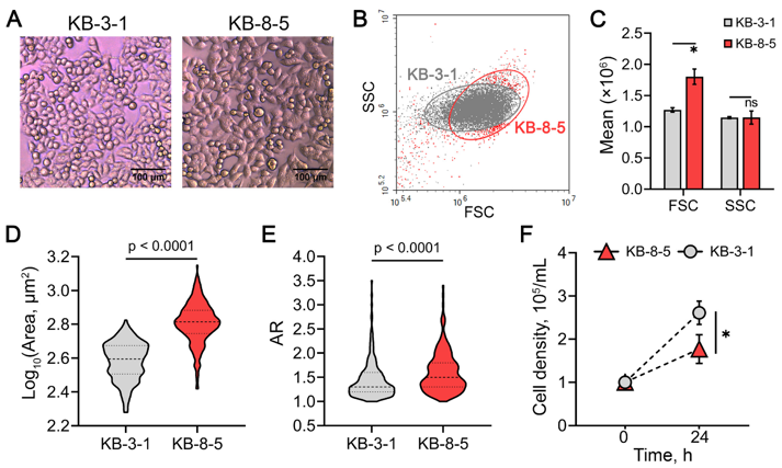
Identification of Key Biological Processes Associated with Chemoresistance Core Genes
Doxorubicin (DOX) is a widely used chemotherapeutic agent, but its efficacy is often limited by cancer cell resistance. Although multiple DOX resistance mechanisms have been characterized, the global transcriptomic alterations underlying this phenomenon remain poorly understood. The aim of this work was to determine whether a common transcriptional response associated with DOX desensitization exists across tumor cells of different origins and to identify the core elements of this response.
We performed an integrated bioinformatics analysis, including: analysis of independent transcriptomic datasets (comparing DOX-resistant neuroblastoma, breast, and cervical carcinoma cells to their DOX-sensitive counterparts), functional annotation of differentially expressed genes, reconstruction and topology analysis of gene networks, text mining, and survival analysis.
We showed that DOX resistance in cancer cells is associated with cytoskeletal reorganization, modulation of cell adhesion, cholesterol biosynthesis, and dysregulation of mTORC1, Wnt, and Gβγ signaling pathways. Experimental validation in DOX-resistant KB-8-5 cervical carcinoma cells and their sensitive counterparts (KB-3-1) confirmed enhanced cellular adhesion and reduced intracellular cholesterol levels associated with chemoresistance, supporting our in-silico findings.
Ask a Question
Write your own review
- You May Also Need

Description: Uterine adenosquamous carcinoma. Said CEA and CA125 producing. Cell growth is slow.

Description: Established from tumor tissue from a 77-year-old woman with recurrence of endometrial carcinoma (adenomatous, partly papillary, grade G3) in 1990; described as forming heterotransplantable tumors in ...

Description: Glassy cell carcinoma. TA-4, CA125, neuron-specific enolase producing.
Description: The cells possess alpha keratin, well defined junctional complexes, tonofilaments and surface microvilli.

- Adipose Tissue-Derived Stem Cells
- Human Neurons
- Mouse Probe
- Whole Chromosome Painting Probes
- Hepatic Cells
- Renal Cells
- In Vitro ADME Kits
- Tissue Microarray
- Tissue Blocks
- Tissue Sections
- FFPE Cell Pellet
- Probe
- Centromere Probes
- Telomere Probes
- Satellite Enumeration Probes
- Subtelomere Specific Probes
- Bacterial Probes
- ISH/FISH Probes
- Exosome Isolation Kit
- Human Adult Stem Cells
- Mouse Stem Cells
- iPSCs
- Mouse Embryonic Stem Cells
- iPSC Differentiation Kits
- Mesenchymal Stem Cells
- Immortalized Human Cells
- Immortalized Murine Cells
- Cell Immortalization Kit
- Adipose Cells
- Cardiac Cells
- Dermal Cells
- Epidermal Cells
- Peripheral Blood Mononuclear Cells
- Umbilical Cord Cells
- Monkey Primary Cells
- Mouse Primary Cells
- Breast Tumor Cells
- Colorectal Tumor Cells
- Esophageal Tumor Cells
- Lung Tumor Cells
- Leukemia/Lymphoma/Myeloma Cells
- Ovarian Tumor Cells
- Pancreatic Tumor Cells
- Mouse Tumor Cells