
- Specification
- Background
- Scientific Data
- Q & A
- Customer Review
HCA-46 (Cellosaurus CVCL_2468) is a well-characterized human colorectal adenocarcinoma cell line derived from a sigmoid colon tumour removed from a 53-year-old female patient. It is epithelial and forms an adherent monolayer in conventional cell culture conditions. Cultures are commonly cultured in DMEM medium supplemented with 2 mM L-glutamine and 10% fetal bovine serum (FBS) at 37°C with 5% CO₂. Morphologically, the cells have epithelial-like characteristics, including tight connections and microvilli, indicating colonic epithelial origin. HCA-46 has been genetically linked to beta-catenin mutations, which are a prevalent molecular hallmark in colorectal cancer. HCA-46 is a commonly used in vitro model for studying colorectal cancer cell biology, signaling pathways, epithelial-mesenchymal transition, and preclinical drug screening for anticancer therapies. Its steady genotype and phenotype make it an excellent tool for researching colorectal tumor growth and treatment response mechanisms.
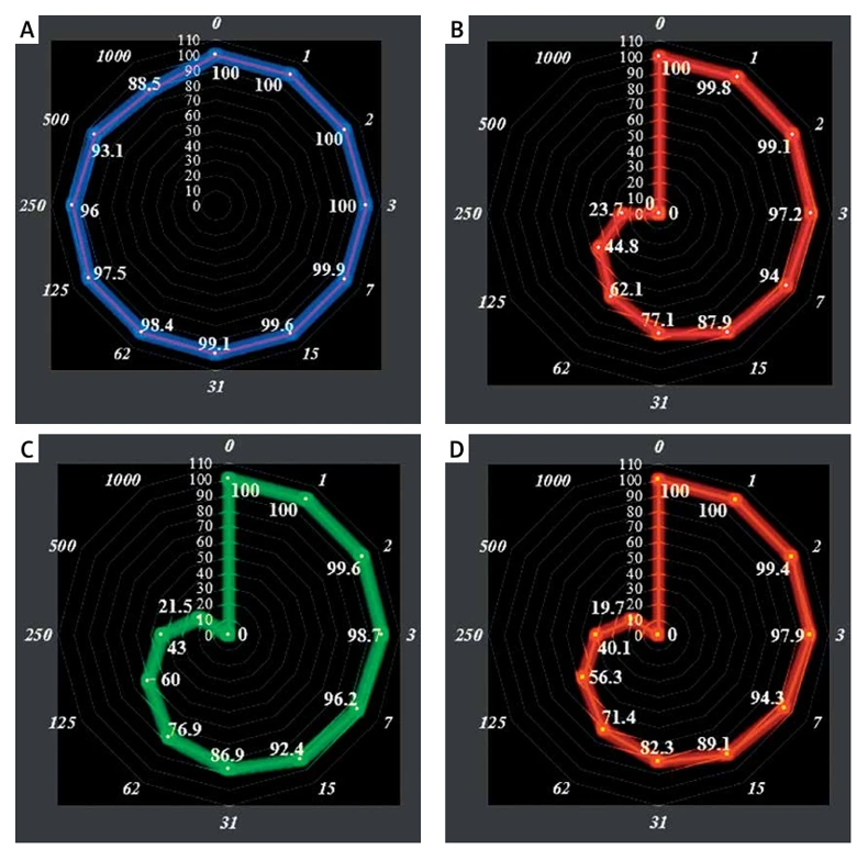
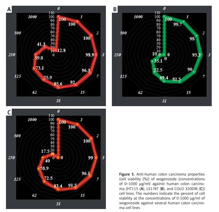
Wogonoside: Anti-Human Colon Cancer Activities and Survey of HMG-Coa Reductase Inhibition Properties with Molecular Modeling
HMG-CoA reductase is a major therapeutic target for cholesterol-lowering medications, and natural compounds with inhibitory potential present promising alternatives for cancer therapy. The objective of this study was to examine wogonoside, a flavonoid compound, as a potential inhibitor of HMG-CoA reductase and to evaluate its efficacy against colon carcinoma.
They used molecular docking and in vitro cytotoxicity tests to see how well wogonoside binds to six colon cancer cell lines and how effectively it might work as a treatment. They did MTT analysis on normal HUVEC cells and malignant colon cell lines (GP5d, MDST8, HCA-46, HT115, LS174T, COLO 320DM) for 48 hours. They used different amounts of wogonoside (Fig. 1 and 2). Wogonoside decreased the viability of malignant colon cell lines in a dose-dependent manner, with IC₅₀ values ranging from 71 to 382 µg/ml (LS174T: 71, GP5d: 105, COLO 320DM: 183, HCA-46: 173, MDST8: 198, HT115: 382). Notably, normal HUVEC cells maintain viability even at 1000 µg/ml (Fig. 1 and 2), indicating selective cytotoxicity toward colon cancer cells.
Ask a Question
Write your own review

Description: Established from an adenocarcinoma located in the ascending colon of a 68 year-old male patient. The adenocarcinoma was well differentiated and determined to be Dukes' stage B. Imperial College ...

Description: This is one cell line out of a series of colon carcinoma cell lines established by PD Dr. Michael Linnebacher.
Description: Rectal carcinoma from a Japanese patient. Cell growth is slow.
Description: The C80 cell line was established from a 69-year old male patient with a moderately well differentiated adenocarcinoma of the rectum classified as Dukes' stage D.
Description: RKO is a poorly differentiated colon carcinoma cell line developed by Michael Brattain.
- Adipose Tissue-Derived Stem Cells
- Human Neurons
- Mouse Probe
- Whole Chromosome Painting Probes
- Hepatic Cells
- Renal Cells
- In Vitro ADME Kits
- Tissue Microarray
- Tissue Blocks
- Tissue Sections
- FFPE Cell Pellet
- Probe
- Centromere Probes
- Telomere Probes
- Satellite Enumeration Probes
- Subtelomere Specific Probes
- Bacterial Probes
- ISH/FISH Probes
- Exosome Isolation Kit
- Human Adult Stem Cells
- Mouse Stem Cells
- iPSCs
- Mouse Embryonic Stem Cells
- iPSC Differentiation Kits
- Mesenchymal Stem Cells
- Immortalized Human Cells
- Immortalized Murine Cells
- Cell Immortalization Kit
- Adipose Cells
- Cardiac Cells
- Dermal Cells
- Epidermal Cells
- Peripheral Blood Mononuclear Cells
- Umbilical Cord Cells
- Monkey Primary Cells
- Mouse Primary Cells
- Breast Tumor Cells
- Colorectal Tumor Cells
- Esophageal Tumor Cells
- Lung Tumor Cells
- Leukemia/Lymphoma/Myeloma Cells
- Ovarian Tumor Cells
- Pancreatic Tumor Cells
- Mouse Tumor Cells