COLO-704
Cat.No.: CSC-C0323
Species: Homo sapiens (Human)
Source: Ascites Metastasis
Morphology: grows as clusters and single cells in suspension
Culture Properties: suspension
- Specification
- Background
- Scientific Data
- Q & A
- Customer Review
Immunology: cytokeratin -, cytokeratin-7 -, cytokeratin-8 -, cytokeratin-17 -, cytokeratin-18 -, desmin -, endothel -, GFAP -, HMB-45 -, neurofilament -, vimentin +
Virus
COLO-704 is a human ovarian adenocarcinoma cell line established in 1986 from ascites of a 46-year-old Caucasian female patient with colon metastasis. The cells show typical morphological epithelial-like features with well-defined cell contours and a high nuclear-cytoplasmic ratio. This cell line grows in suspension, distributing as single cells or small clusters in RPMI 1640 medium supplemented with 10% FBS, and requires subculturing at a 1:2-1:6 ratio twice weekly under 37°C and 5% CO₂ conditions. On a molecular level, the cell line expresses oncogenes nuclear v-myc and chromosomal v-raf, shows env gp70 antigen on the cell surface and contains 380 mutated genes related to apoptosis, metabolism and signal transduction (e.g. AATK, ACOX3). The cell line serves as an ovarian cancer research tool to study metastasis mechanisms alongside drug screening of chemotherapeutic and targeted drugs and validation of molecular biomarkers that indicate cross organ metastatic potential.
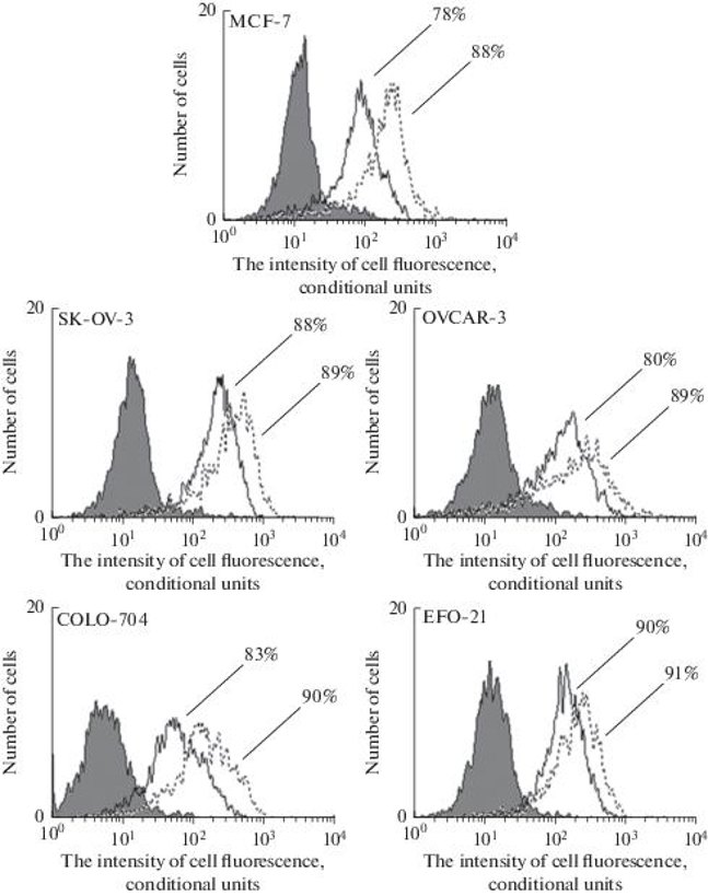
Comparative Immunofluorescent Analysis of Estrogen Receptor Beta Expression in Ovarian and Breast Cancer Cell Lines
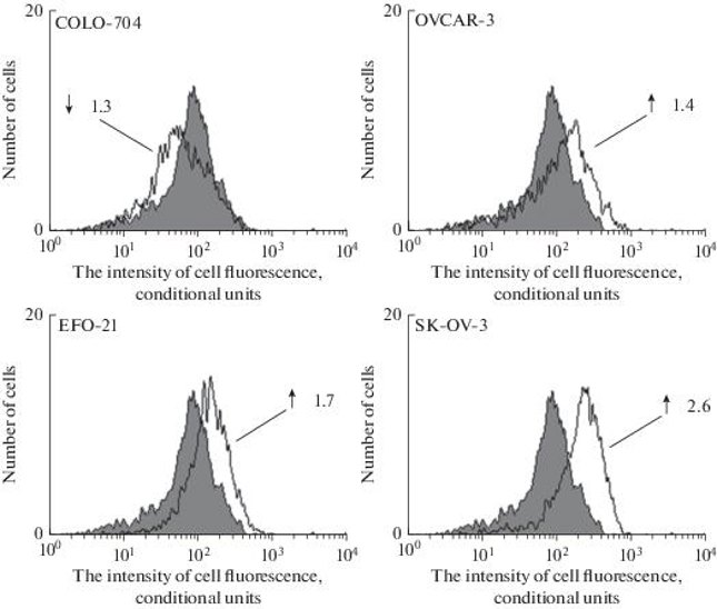
Although the target of tamoxifen in breast cancer is estrogen receptor α (ERα), the target of tamoxifen in ovarian cancer is still unknown. Estrogen receptor β (ERβ) is a cell signaling and proliferation transcription factor, and its expression level in ovarian cancer tissues is higher than the level of ERα. But data on the comparison of ERβ expression in ovarian and breast cancer cell lines are absent. Therefore, Bougsh et al. are devoted to the quantitative evaluation of ERβ expression in ovarian cancer cell lines (COLO-704, OVCAR-3, EFO-21, SK-OV-3) and its comparison with the breast cancer cell line MCF-7 using immunofluorescence and flow cytometry. In general, ERβ expression was observed in all cell cultures, and no significant differences in the level of this receptor expression between MCF-7 breast cancer cell line and ovarian cancer cell cultures were found. The expression of ERβ was 88% in MCF-7 cells, and 89, 88, 91 and 90% in SK-OV-3, OVCAR-3, EFO-21 and COLO-704 cells, respectively (Fig. 1). Thus, all studied cell cultures had high levels of ERβ expression. But the degree of expression of this receptor is different. The typical result of one experiment is shown in Fig. 2. Only the ovarian cancer cell line COLO-704 had the degree of ERβ expression similar to MCF-7, being only 1.3 times lower. The SK-OV-3, EFO-21 and OVCAR-3 cell lines had 2.7, 1.4 and 1.9 times higher ERβ expression levels than MCF-7, respectively.
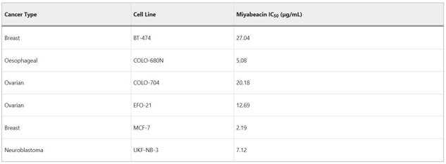
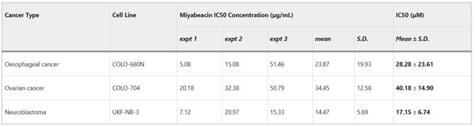
Miyabeacin: A New Cyclodimer Presents a Potential Role for Willow in Cancer Therapy
Willow (Salix spp.) is a well-known source of medicinal compounds, including salicin, the precursor to aspirin. Here, Ward et al. report the isolation and structure determination of a cyclodimeric salicinoid, miyabeacin, from S. miyabeana and S. dasyclados, and its anti-cancer activity. They screened this compound for its anti-cancer activity. Miyabeacin was tested on a range of cancer cell lines including MYCN-amplified neuroblastoma (UKF-NB-3) and its vincristine resistant sub-line (UKF-NB-3rVCR10). At a concentration of 20 µg/mL, it reduced the number of viable cells to 0% and 4.22 ± 2.89% in UKF-NB-3 and UKF-NB-3rVCR10, respectively, after 120 hours. They also derived IC50 values for neuroblastoma (UKF-NB-3), breast (BT-474, MCF-7), oesophageal (COLO-680N) and ovarian cancers (COLO-704, EFO-21) (Fig. 3). These were in the range 2.19-27.04 µg/mL. Fully replicated IC50 data for three selected cell lines are presented in Figure 4. The mean IC50 values were 14.47 ± 5.69 µg/mL (UKF-NB-3), 23.87 ± 19.93 µg/mL (COLO-680N) and 34.45 ± 12.58 µg/mL (COLO-704).
Ask a Question
Write your own review
- You May Also Need
Description: established from ovary of a 47-year-old female with ovarian endometrioid adenocarcinoma

Description: The A2780 human ovarian cancer cell line was established from tumour tissue from an untreated patient. Cells grow as a monolayer and in suspension in spinner cultures. A2780 is the parent line to the ...

Description: Japanese ovarian serous adenocarcinoma. Said CA125 producing. Cell growth is slow.

Description: Established post-hysterectomy from the tumor tissue of a 65-year-old Japanese woman with malignant ovarian carcinoma (stage: FIGO IIIc; histology: poorly differentiated serous papillary ...

- Adipose Tissue-Derived Stem Cells
- Human Neurons
- Mouse Probe
- Whole Chromosome Painting Probes
- Hepatic Cells
- Renal Cells
- In Vitro ADME Kits
- Tissue Microarray
- Tissue Blocks
- Tissue Sections
- FFPE Cell Pellet
- Probe
- Centromere Probes
- Telomere Probes
- Satellite Enumeration Probes
- Subtelomere Specific Probes
- Bacterial Probes
- ISH/FISH Probes
- Exosome Isolation Kit
- Human Adult Stem Cells
- Mouse Stem Cells
- iPSCs
- Mouse Embryonic Stem Cells
- iPSC Differentiation Kits
- Mesenchymal Stem Cells
- Immortalized Human Cells
- Immortalized Murine Cells
- Cell Immortalization Kit
- Adipose Cells
- Cardiac Cells
- Dermal Cells
- Epidermal Cells
- Peripheral Blood Mononuclear Cells
- Umbilical Cord Cells
- Monkey Primary Cells
- Mouse Primary Cells
- Breast Tumor Cells
- Colorectal Tumor Cells
- Esophageal Tumor Cells
- Lung Tumor Cells
- Leukemia/Lymphoma/Myeloma Cells
- Ovarian Tumor Cells
- Pancreatic Tumor Cells
- Mouse Tumor Cells