
MKPL-1
Cat.No.: CSC-C6762J
Species: Homo sapiens (Human)
Source: Bone Marrow
Morphology: Primitive Blast
- Specification
- Background
- Scientific Data
- Q & A
- Customer Review
The MKPL-1 is a human leukemic cell line. The cells are isolated from the peripheral blood of a 36-year-old male patient with acute megakaryoblastic leukemia (AMKL), a rare and often aggressive subtype of acute myeloid leukemia (AML). The MKPL-1 cell line, as such, provides an in vitro model for AMKL. The MKPL-1 cells, as cultured in vitro, display a blast-like morphology and show growth in suspension as single cells or small aggregates. The doubling time is approximately 24-48 hours. A key morphologic and functional feature of this cell line is the ability of the cells to demonstrate features of megakaryocytic differentiation upon treatment with specific pharmacological stimuli, such as phorbol esters (PMA). This can be assessed both by morphological changes (e.g. expansion of cytoplasm, protrusions) and an increased expression of megakaryocytic surface markers such as CD41, and CD61.
Functionally, MKPL-1 is an important model to study the pathophysiology of AMKL in particular and to do research involving aberrant megakaryopoiesis and leukemogenesis. Its main applications are for drug screens for novel anti-leukemic compounds, drug resistance studies, as well as differentiation therapy approaches. The cell line has also been used for molecular research looking at the role of specific genetic alterations in leukemogenesis, such as the CBFA2T3-GLIS2 fusion oncogene which is often associated with poor prognosis in pediatric AMKL.
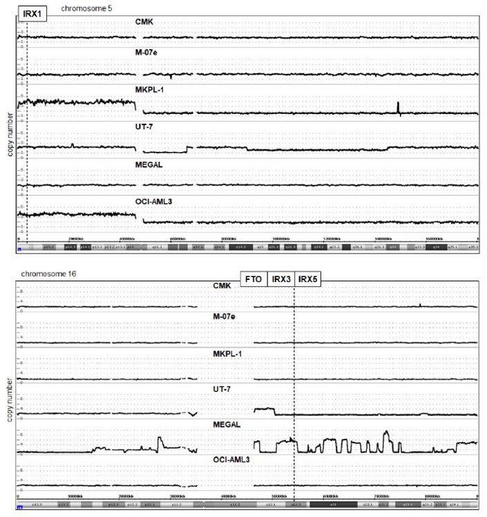
Chromosomal and Genomic Analyses of the Gene Loci for IRX1, IRX3, and IRX5 in AML
Homeobox genes encode transcription factors crucial for development, including hematopoiesis. Recently, the lymphoid TALE-code was constructed, detailing TALE homeobox gene expression in early hematopoiesis. Building on the lymphoid TALE-code, Nagel et al. constructed the myeloid TALE-code to cover the entire hematopoietic system. Then, they investigated the role of TALE homeobox genes, particularly IRX1, IRX3, and IRX5, in myeloid differentiation and acute myeloid leukemia (AML).
To determine if chromosomal rearrangements activate IRX genes in AML, they reviewed published karyotypes of relevant cell lines and identified i(5)(p10) in OCI-AML3 and MKPL-1 and del(16)(q13q23) in MEGAL as potential activating alterations. They then performed genomic profiling on AML cell lines CMK, M-07e, MKPL-1, UT-7, MEGAL, and OCI-AML3, focusing on chromosomes 5 and 16 (Fig. 1). IRX1 (5p15) is duplicated in OCI-AML3 and MKPL-1, consistent with isochromosome formation for the short arm. However, OCI-AML3 is IRX1-negative, suggesting this duplication does not activate IRX1. In contrast, IRX3 and IRX5 at 16q12 are significantly amplified in MEGAL, with several amplicons covering the entire long arm of chromosome 16. RNA-seq data for the nearby FTO locus, which regulates IRX3/IRX5 activity, showed enhanced expression in MEGAL. This genomic amplification likely drives IRX3/IRX5 activation in MEGAL. RT-PCR confirmed no CBFb-MYH11 fusion gene formation via inv(16)(p13q22) in MEGAL.
Ask a Question
Write your own review
- You May Also Need

Description: Established in 2007 from the bone marrow mononuclear cells of an 82-year-old Japanese man with diffuse large B-cell lymphoma in the leukemic phase

Description: Established from the bone marrow of a 28-year-old man who developed the terminal leukemic phase of lymphosarcoma in 1976

Description: This cell line was derived from the bone marrow aspirate of a 59 year old male with erythroleukemia that became acute myelogenous leukaemia.The cells form colonies in soft-agar in the presence of ...

Description: Established from the pleural effusion of a 24-year-old woman with recurrent anaplastic large cell lymphoma (ALCL); cells were described to clonally derive from T-lineage lymphoid cells and to be ...

Description: Established from a 37-year-old man at second (refractory/terminal) relapse of Hodgkin lymphoma (nodular sclerosing -> lymphocyte depleted/stage IIISA -> stage IV) after both combined chemo- and ...

Description: Established from the peripheral blood of a 10-year-old Caucasian boy with acute lymphoblastic leukemia (pre B-ALL) at diagnosis in 1993
- Adipose Tissue-Derived Stem Cells
- Human Neurons
- Mouse Probe
- Whole Chromosome Painting Probes
- Hepatic Cells
- Renal Cells
- In Vitro ADME Kits
- Tissue Microarray
- Tissue Blocks
- Tissue Sections
- FFPE Cell Pellet
- Probe
- Centromere Probes
- Telomere Probes
- Satellite Enumeration Probes
- Subtelomere Specific Probes
- Bacterial Probes
- ISH/FISH Probes
- Exosome Isolation Kit
- Human Adult Stem Cells
- Mouse Stem Cells
- iPSCs
- Mouse Embryonic Stem Cells
- iPSC Differentiation Kits
- Mesenchymal Stem Cells
- Immortalized Human Cells
- Immortalized Murine Cells
- Cell Immortalization Kit
- Adipose Cells
- Cardiac Cells
- Dermal Cells
- Epidermal Cells
- Peripheral Blood Mononuclear Cells
- Umbilical Cord Cells
- Monkey Primary Cells
- Mouse Primary Cells
- Breast Tumor Cells
- Colorectal Tumor Cells
- Esophageal Tumor Cells
- Lung Tumor Cells
- Leukemia/Lymphoma/Myeloma Cells
- Ovarian Tumor Cells
- Pancreatic Tumor Cells
- Mouse Tumor Cells