
Immortalized Mouse Progenitor-Like Melanocytes (iMC23)
Cat.No.: CSC-I9233L
Species: Mus musculus
Source: Skin
Morphology: Polygonal
Culture Properties: Adherent
- Specification
- Background
- Scientific Data
- Q & A
- Customer Review
Note: Never can cells be kept at -20 °C.
2) Tyrosine enzymatic assays determined presence of tyrosine;
3) Western blot analysis for SV40 large T antigen presence
Immortalized Mouse Progenitor-Like Melanocytes (iMC23) is a non-tumorigenic immortalized murine cell line originally isolated from mouse (C57BL/6) skin, and immortalized using the SV40 large T antigen viral transduction. They display a progenitor-like melanocytic phenotype, and expression of melanocytic progenitor markers like c-kit, Pax3, Sox10 and MITF-m. iMC23 grow as an adherent cell line in culture with a polygonal morphology. They are selected using hygromycin resistance.
Despite being non-pigmented as is, iMC23 can be differentiated with dexamethasone to a mature melanocyte phenotype, with expression of melanocyte markers such as HMB45 positivity and low tyrosinase activity. They have been used as a tool to study melanogenesis in vitro and also molecular regulation of melanocyte maturation. Specifically, iMC23 has been utilized to study pathways of melanocyte differentiation, like the Wnt/β-catenin pathway. Overexpression of Wnt10b can cause these progenitor-like melanocytes to differentiate to pigmented, active melanocytes with upregulated tyrosinase activity.
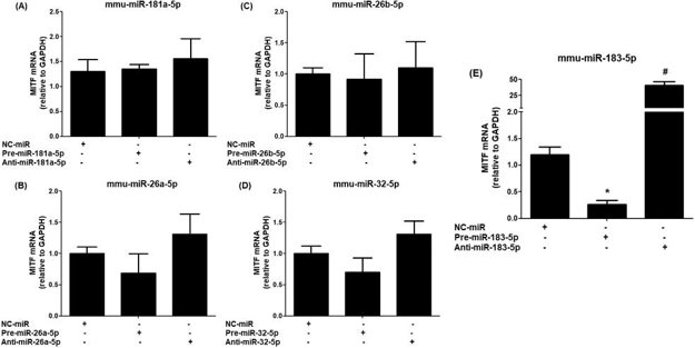
Determination of Correlation of MITF Expression with Dysregulated miRNAs
MITF is an important regulator for melanocytes, and miRNAs are important for various biological functions. In this study, AI Robaee et al. induced the depigmentation of C57BL/6 mice and used bioinformatics to screen for miRNAs that target the MITF 3'UTR. Then, the iMC23 mouse melanocyte cell line was used to validate the bioinformatics and LS-VtM, NLS-VtM, and NS-CM data. Mimics or inhibitors of mmu-miR-181a-5p, mmu-miR-26a-5p, mmu-miR-26b-5p, mmu-miR-32-5p and mmu-miR-183-5p were transfected in iMC23 cells and the MITF mRNA levels were measured. In addition, NC-miR was transfected as a negative control. As shown in Fig. 1A-D, mimics or inhibitors transfection of mmu-miR-181a-5p, mmu-miR-26a-5p, mmu-miR-26b-5p, and mmu-miR-32-5p, respectively, did not change the MITF mRNA expression. In contrast, pre-miR-183-5p transfection significantly downregulated MITF mRNA and anti-miR-183-5p transfection significantly upregulated MITF mRNA (Fig. 1E). These data show that among all the screened miRNAs, only mmu-miR-183-5p regulates MITF mRNA expression in iMC23 melanocytes.
Ask a Question
Write your own review
Description: Immortalized Mouse Primary Artery Fibroblasts-GFP provided by Creative Bioarray have been developed by immortalizing mouse artery fibroblasts with SV40 Large T antigen and transfecting with tGFP. The ...

Description: Immortalized MHC II -/- Mouse Macrophage Cells (C2D) is derived from the knockout mice negative for MHCII molecule. They are stable in culture and survived through crisis in which they spontaneously ...
Description: The bone morphogenetic proteins (BMPs) produced by calvarial cells are crucial in osteoblastic differentiation and bone regeneration, however the limited lifespan of primary cells diminishes their ...

Description: The Immortalized Mouse Atrioventricular Cushion Mesenchymal Cells (tsA58-AVM) were derived from atrioventricular (AV) cushions of H-2Kb -tsA58 embryos at E9.5 and were conditionally immortalized in ...
Description: IDG-SW3 represent a non-homogenous population progressing from early osteoblasts to late osteocytic. These cells express functional SV40 large T antigen that is induced in the presence of IFN γ under ...
Description: The Immortalized Mouse Spleen Dendritic Cell (SRDC) line is functionally and phenotypically similar to dendritic cells; specifically, in its antigen presentation, T cell priming, and dendritic cell ...
- Adipose Tissue-Derived Stem Cells
- Human Neurons
- Mouse Probe
- Whole Chromosome Painting Probes
- Hepatic Cells
- Renal Cells
- In Vitro ADME Kits
- Tissue Microarray
- Tissue Blocks
- Tissue Sections
- FFPE Cell Pellet
- Probe
- Centromere Probes
- Telomere Probes
- Satellite Enumeration Probes
- Subtelomere Specific Probes
- Bacterial Probes
- ISH/FISH Probes
- Exosome Isolation Kit
- Human Adult Stem Cells
- Mouse Stem Cells
- iPSCs
- Mouse Embryonic Stem Cells
- iPSC Differentiation Kits
- Mesenchymal Stem Cells
- Immortalized Human Cells
- Immortalized Murine Cells
- Cell Immortalization Kit
- Adipose Cells
- Cardiac Cells
- Dermal Cells
- Epidermal Cells
- Peripheral Blood Mononuclear Cells
- Umbilical Cord Cells
- Monkey Primary Cells
- Mouse Primary Cells
- Breast Tumor Cells
- Colorectal Tumor Cells
- Esophageal Tumor Cells
- Lung Tumor Cells
- Leukemia/Lymphoma/Myeloma Cells
- Ovarian Tumor Cells
- Pancreatic Tumor Cells
- Mouse Tumor Cells