

Immortalized Mouse Lymphatic Endothelial Cells-SV40
Cat.No.: CSC-I2192Z
Species: mouse
Morphology: Polygonal
Culture Properties: Adherent
- Specification
- Background
- Scientific Data
- Q & A
- Customer Review
free from contaminations (bacteria incl. mycoplasma, fungi, HIV, HAV, HBV, HCV, Parvo-B19) and cross-contaminations
Note: Never can cells be kept at -20°C.
Immortalized mouse lymphatic endothelial cells (iMLECs), typically generated by stable expression of the SV40 large T antigen, provide a powerful and practical model for studying lymphatic biology. A central advantage of iMLECs is their faithful retention of lineage-defining lymphatic markers. They robustly express the master transcription factor Prox1, the surface glycoprotein podoplanin, the hyaluronan receptor LYVE-1, and the vascular endothelial growth factor receptor VEGFR-3, while lacking the blood endothelial marker von Willebrand factor. Functionally, these cells mount potent proliferative, migratory, and tube-formation responses to VEGF-C, recapitulating key lymphangiogenic steps. They also maintain active acetylated low-density lipoprotein uptake and upregulate adhesion molecules under inflammatory stimuli, demonstrating preserved endothelial cell functions.
Compared with primary LECs, iMLECs offer a homogeneous, genetically stable population that eliminates donor-to-donor variability and drastically reduces animal use. Their unlimited expansion supports long-term studies, high-throughput drug screening, and sophisticated genetic manipulation, including lentiviral transduction and CRISPR/Cas9 editing. Critically, the combination of stable antigenic profiles and consistent functional responsiveness makes them a highly reproducible platform for dissecting lymphangiogenic signaling, evaluating anti-lymphangiogenic therapies, and modeling lymphatic involvement in tumor metastasis and chronic inflammation.
LEC–Cancer Cell Adhesion Induces Reciprocal Changes in Actin Reorganization
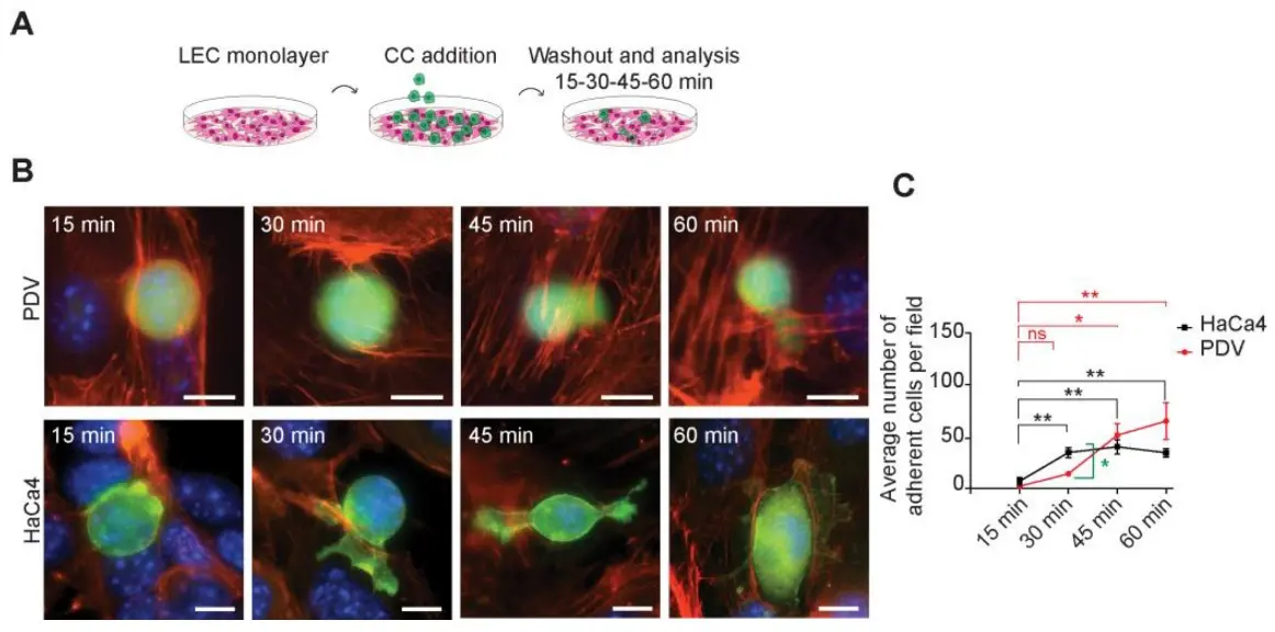
Cell adhesion in carcinogenesis is positively and negatively regulated, enabling cancer cells to attach to other cells and the extracellular matrix, favoring their permanence at the cancer stem cells (CSCs) niche or their migration and dissemination to distant sites. The actin cytoskeleton plays a critical role in cell adhesion and migration. Therefore, this study sought to identify the occurrence of changes in actin organization at sites of heterotypic cell–cell interaction between lymphatic endothelial cells (LECs) and the cancer cell lines.
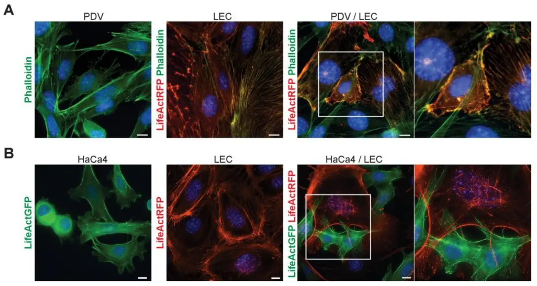
For this purpose, we used the immortalized mouse LEC line and two mouse skin squamous carcinoma cell lines, PDV and HaCa4, both of which express the CSC marker SOX2. In individual cell cultures, LEC cells grew as compact adhesive cell monolayers, displaying a well-defined cortical actin network forming a honeycomb pattern with radial bundles of actin fibers at cell–cell contact sites. On the other hand, epithelial PDV and the epithelioid HaCa4 cells established cell–cell contacts with a polarized actin cytoskeleton organization.
Upon co-culture, we observed distinct morphological changes in both LECs and cancer cells. LEC cells exhibited a more elongated organization and no longer formed a close monolayer. Instead, they organized into an open network, establishing contact with cancer cells. The actin cytoskeleton of LEC and cancer cells appeared reorganized, and areas of directed actin membrane protrusions were evident at heterotypic cell adhesion sites, along with the presence of actin bundles and lamellipodia membrane protrusions. These results underscore the existence of productive adhesive interactions involving the actin cytoskeleton between LECs and cancer cells.
Ask a Question
Write your own review
Description: Immortalized Cynomolgus Monkey Pulmonary Artery Smooth Muscle Cells-GFP provided by Creative Bioarray have been developed by immortalizing cynomolgus monkey pulmonary artery smooth muscle cells with ...
Description: Immortalized Monkey Spleen Fibroblasts-SV40T were developed from monkey tissues transduced with a lentiviral expression vector containing the SV40T gene. The cell line was continuously cultured for ...

Description: Immortalized Monkey Bronchial Fibroblasts-GFP provided by Creative Bioarray have been developed by immortalizing monkey bronchial fibroblasts with SV40 Large T antigen and transfecting with tGFP. The ...
Description: Immortalized Canine Kidney Fibroblasts-SV40 have been obtained immortalizing Canine Kidney Fibroblasts with Lenti-SV40 Lentivirus. Immortalized cells were controlled passaging side by side with the ...
Description: Immortalized Monkey Primary Aortic Endothelial Cells-GFP provided by Creative Bioarray have been developed by immortalizing primary monkey aortic endothelial cells with SV40 Large T antigen and ...
Description: Immortalized Monkey Pancreatic Epithelial Cells-SV40T were developed from monkey tissues transduced with a lentiviral expression vector containing the SV40T gene. The cell line was continuously ...
- Adipose Tissue-Derived Stem Cells
- Human Neurons
- Mouse Probe
- Whole Chromosome Painting Probes
- Hepatic Cells
- Renal Cells
- In Vitro ADME Kits
- Tissue Microarray
- Tissue Blocks
- Tissue Sections
- FFPE Cell Pellet
- Probe
- Centromere Probes
- Telomere Probes
- Satellite Enumeration Probes
- Subtelomere Specific Probes
- Bacterial Probes
- ISH/FISH Probes
- Exosome Isolation Kit
- Human Adult Stem Cells
- Mouse Stem Cells
- iPSCs
- Mouse Embryonic Stem Cells
- iPSC Differentiation Kits
- Mesenchymal Stem Cells
- Immortalized Human Cells
- Immortalized Murine Cells
- Cell Immortalization Kit
- Adipose Cells
- Cardiac Cells
- Dermal Cells
- Epidermal Cells
- Peripheral Blood Mononuclear Cells
- Umbilical Cord Cells
- Monkey Primary Cells
- Mouse Primary Cells
- Breast Tumor Cells
- Colorectal Tumor Cells
- Esophageal Tumor Cells
- Lung Tumor Cells
- Leukemia/Lymphoma/Myeloma Cells
- Ovarian Tumor Cells
- Pancreatic Tumor Cells
- Mouse Tumor Cells