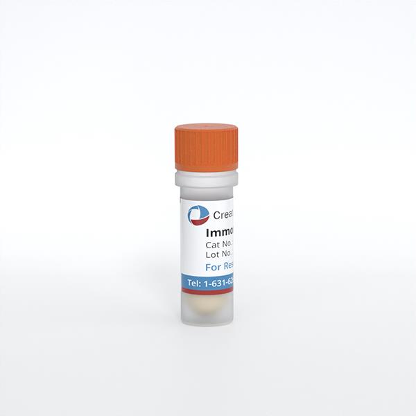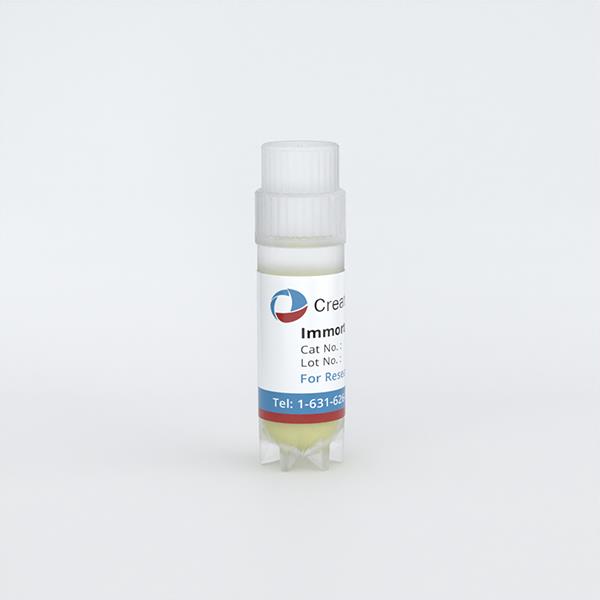
Immortalized Mouse Intestinal Myofibroblasts
Cat.No.: CSC-I9228L
Species: Mus musculus
Source: Colon
Morphology: Spindle-shaped
Culture Properties: Adherent
- Specification
- Q & A
- Customer Review
Cat.No.
CSC-I9228L
Description
Intestinal epithelial cells provide a protective barrier against bacteria, antigens, and toxic substances which enters the body through the luminal tube. The cells are regulated to differentiate and proliferate to establish their protective function. Subepithelial intestinal myofibroblasts have been indicated to have a role in regulating intestinal epithelial cells. The Immortalized Mouse Intestinal Myofibroblasts are isolated from C57BL/6J mouse colonic muscosa and transfected with SV40 large T antigen. It retains primary myofibroblastic phenotypes, and expresses α-smooth muscle actin, vimentin, and type I collagen. Immune response-related proteins, such as TLR-4, CD14, MD2, and IκBα are also detected. Additionally, these cells are reactive to lipopolysaccharide (LPS) treatment, enabling an inflammatory response causing the degradation of IκBα and p38 MAPK phosphorylation. It is a valuable tool in research involving intestinal injury, fibrosis, inflammation, tissue repair, and immune regulation in intestinal tissue.
Species
Mus musculus
Source
Colon
Culture Properties
Adherent
Morphology
Spindle-shaped
Immortalization Method
Serial passaging and transduction with recombinant lentiviruses carrying SV40 Large T antigen
Markers
α-smooth muscle actin, vimentin, type I collagen
Application
For Research Use Only
Storage
Directly and immediately transfer cells from dry ice to liquid nitrogen upon receiving and keep the cells in liquid nitrogen until cell culture needed for experiments.
Note: Never can cells be kept at -20 °C.
Note: Never can cells be kept at -20 °C.
Shipping
Dry Ice.
Recommended Products
CIK-HT003 HT® Lenti-SV40T Immortalization Kit
Quality Control
Western blot and immunofluorescence staining were used to determined cell markers and protein expression.
BioSafety Level
II
Citation Guidance
If you use this products in your scientific publication, it should be cited in the publication as: Creative Bioarray cat no.
If your paper has been published, please click here
to submit the PubMed ID of your paper to get a coupon.
Ask a Question
Write your own review
Related Products
Featured Products
- Adipose Tissue-Derived Stem Cells
- Human Neurons
- Mouse Probe
- Whole Chromosome Painting Probes
- Hepatic Cells
- Renal Cells
- In Vitro ADME Kits
- Tissue Microarray
- Tissue Blocks
- Tissue Sections
- FFPE Cell Pellet
- Probe
- Centromere Probes
- Telomere Probes
- Satellite Enumeration Probes
- Subtelomere Specific Probes
- Bacterial Probes
- ISH/FISH Probes
- Exosome Isolation Kit
- Human Adult Stem Cells
- Mouse Stem Cells
- iPSCs
- Mouse Embryonic Stem Cells
- iPSC Differentiation Kits
- Mesenchymal Stem Cells
- Immortalized Human Cells
- Immortalized Murine Cells
- Cell Immortalization Kit
- Adipose Cells
- Cardiac Cells
- Dermal Cells
- Epidermal Cells
- Peripheral Blood Mononuclear Cells
- Umbilical Cord Cells
- Monkey Primary Cells
- Mouse Primary Cells
- Breast Tumor Cells
- Colorectal Tumor Cells
- Esophageal Tumor Cells
- Lung Tumor Cells
- Leukemia/Lymphoma/Myeloma Cells
- Ovarian Tumor Cells
- Pancreatic Tumor Cells
- Mouse Tumor Cells
Hot Products

