
Immortalized Mouse Hepatocytes-SV40
Cat.No.: CSC-I2205Z
Species: mouse
Morphology: Polygonal
Culture Properties: Adherent
- Specification
- Background
- Scientific Data
- Q & A
- Customer Review
free from contaminations (bacteria incl. mycoplasma, fungi, HIV, HAV, HBV, HCV, Parvo-B19) and cross-contaminations
Note: Never can cells be kept at -20°C.
Immortalized Mouse Hepatocytes-SV40 are derived from C57BL/6 mouse liver and engineered for continuous proliferation via lentiviral transduction of SV40 large T antigen, with transgene expression validated by real-time PCR. These adherent, polygonal cells overcome the fundamental limitation of primary hepatocytes—rapid onset of replicative senescence after the first passage—by maintaining robust growth beyond 20 passages without loss of viability or phenotypic integrity.
Compared to conventional hepatoma lines, this immortalized model retains closer fidelity to native hepatic biology while offering unlimited experimental scalability. The cells provide a renewable, genetically stable platform for investigating hepatocyte biology, hepatic metabolism, stress responses, and disease-associated signaling pathways. Their well-defined C57BL/6 background ensures reproducibility across genetic, transcriptomic, and compound-response studies.
With demonstrated utility in gene expression analysis, drug metabolism, and toxicity screening, Immortalized Mouse Hepatocytes-SV40 bridge the gap between short-lived primary cultures and fully transformed cell lines. They deliver the physiological relevance of hepatocyte-derived models together with the experimental consistency and throughput essential for mechanistic research and preclinical evaluation.
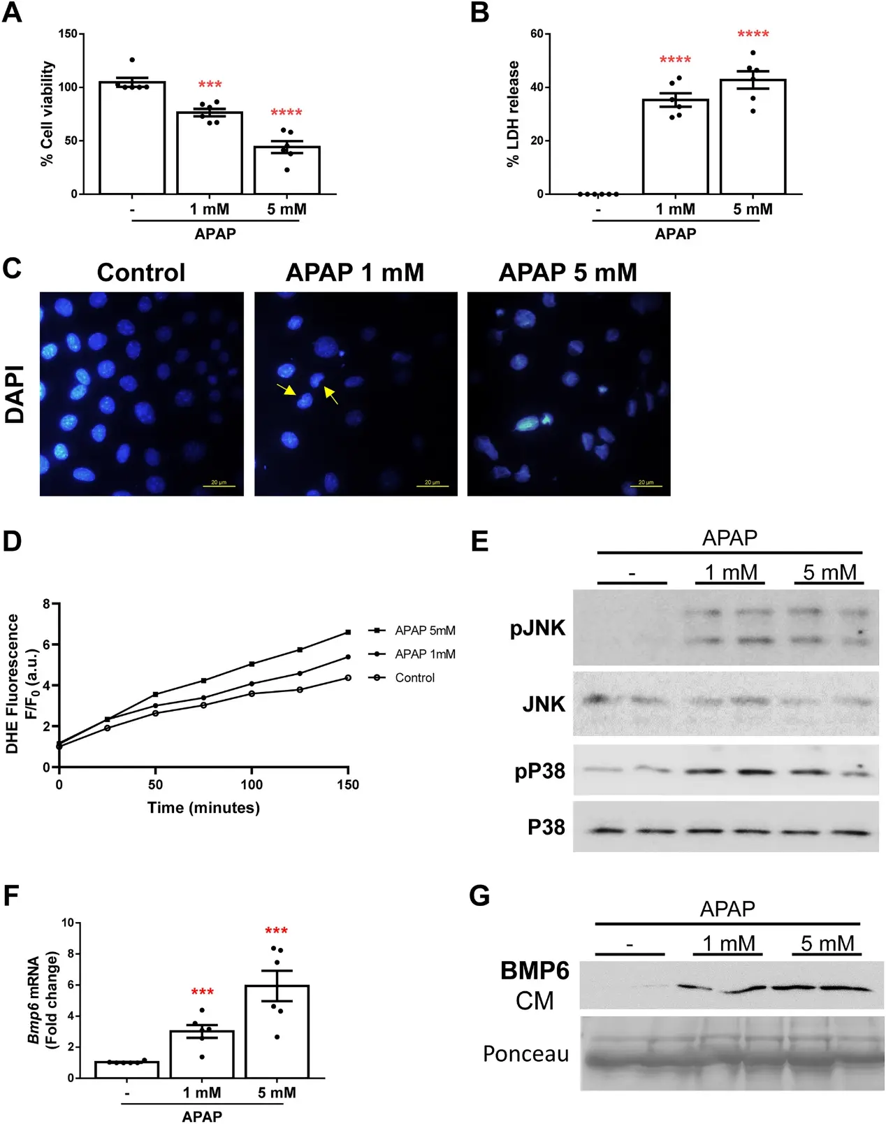
BMP6 Participates in the Molecular Mechanisms Involved in APAP Hepatotoxicity
Given the lack of accurate diagnostic methods of acetaminophen (APAP)-induced acute liver failure (ALF), the search for new biomarkers for its diagnosis is an urgent need. The aim of this study was to evaluate the role of bone morphogenetic protein 6 (BMP6) in APAP-induced ALF progression and its potential value as a biomarker of ALF.
Immortalized neonatal mouse hepatocytes were exposed to APAP (1 mM and 5 mM) for 16 h to assess APAP-induced cell death. As shown in Fig. 1A, we observed a decrease in cell viability in a concentration-dependent manner. Moreover, both APAP concentrations had cytotoxic effects reflected by the release of LDH (Fig. 1B) and presence of apoptotic nuclei (Fig. 1C). Furthermore, we used DHE probe to measure reactive oxygen species (ROS) production in these cells during 2 h after APAP stimulation and we found that APAP-induced ROS (Fig. 1D) and, thus, ROS-mediated JNK and P38 MAPK phosphorylation was also observed (Fig. 1E). Next, we measured Bmp6 mRNA levels and found a significant increase in hepatocytes treated with APAP compared to the controls (Fig. 1F). In addition, BMP6 was detected in the culture media (CM) from cells exposed to APAP (Fig. 1G).
Ask a Question
Write your own review

Description: Immortalized Cynomolgus Monkey Pulmonary Artery Smooth Muscle Cells-GFP provided by Creative Bioarray have been developed by immortalizing cynomolgus monkey pulmonary artery smooth muscle cells with ...

Description: Immortalized Monkey Spleen Fibroblasts-SV40T were developed from monkey tissues transduced with a lentiviral expression vector containing the SV40T gene. The cell line was continuously cultured for ...
Description: Immortalized Monkey Bronchial Fibroblasts-GFP provided by Creative Bioarray have been developed by immortalizing monkey bronchial fibroblasts with SV40 Large T antigen and transfecting with tGFP. The ...
Description: Immortalized Canine Kidney Fibroblasts-SV40 have been obtained immortalizing Canine Kidney Fibroblasts with Lenti-SV40 Lentivirus. Immortalized cells were controlled passaging side by side with the ...
Description: Immortalized Monkey Primary Aortic Endothelial Cells-GFP provided by Creative Bioarray have been developed by immortalizing primary monkey aortic endothelial cells with SV40 Large T antigen and ...
Description: Immortalized Monkey Pancreatic Epithelial Cells-SV40T were developed from monkey tissues transduced with a lentiviral expression vector containing the SV40T gene. The cell line was continuously ...
- Adipose Tissue-Derived Stem Cells
- Human Neurons
- Mouse Probe
- Whole Chromosome Painting Probes
- Hepatic Cells
- Renal Cells
- In Vitro ADME Kits
- Tissue Microarray
- Tissue Blocks
- Tissue Sections
- FFPE Cell Pellet
- Probe
- Centromere Probes
- Telomere Probes
- Satellite Enumeration Probes
- Subtelomere Specific Probes
- Bacterial Probes
- ISH/FISH Probes
- Exosome Isolation Kit
- Human Adult Stem Cells
- Mouse Stem Cells
- iPSCs
- Mouse Embryonic Stem Cells
- iPSC Differentiation Kits
- Mesenchymal Stem Cells
- Immortalized Human Cells
- Immortalized Murine Cells
- Cell Immortalization Kit
- Adipose Cells
- Cardiac Cells
- Dermal Cells
- Epidermal Cells
- Peripheral Blood Mononuclear Cells
- Umbilical Cord Cells
- Monkey Primary Cells
- Mouse Primary Cells
- Breast Tumor Cells
- Colorectal Tumor Cells
- Esophageal Tumor Cells
- Lung Tumor Cells
- Leukemia/Lymphoma/Myeloma Cells
- Ovarian Tumor Cells
- Pancreatic Tumor Cells
- Mouse Tumor Cells