

Immortalized Mouse Dermal Lymphatic Endothelial Cells-SV40
Cat.No.: CSC-I2191Z
Species: mouse
Morphology: Polygonal
Culture Properties: Adherent
- Specification
- Background
- Scientific Data
- Q & A
- Customer Review
free from contaminations (bacteria incl. mycoplasma, fungi, HIV, HAV, HBV, HCV, Parvo-B19) and cross-contaminations
Note: Never can cells be kept at -20°C.
Immortalized mouse dermal lymphatic endothelial cells (LECs) are generated by transducing primary murine dermal LECs with a retroviral or lentiviral vector encoding the simian virus 40 (SV40) large T antigen. The distinct advantages of this cell line are threefold. First, indefinite proliferation and scalability – SV40‑immortalised LECs can be propagated beyond 30 passages with consistent doubling times, ensuring a reliable and abundant supply for long‑term and high‑throughput studies, eliminating the batch‑to‑batch variability inherent to primary isolates. Second, faithful retention of lymphatic identity – these cells maintain characteristic cobblestone morphology and express the full panel of definitive lymphatic markers, including podoplanin, Prox‑1 (a master transcriptional regulator), VEGFR‑3 (Flt‑4), and LYVE‑1, while remaining negative for blood vascular markers such as CD34. This molecular signature is stably maintained over extended culture, confirming their specialized lineage. Third, functional competence – immortalized LECs form capillary‑like tubular networks on Matrigel, actively migrate in response to VEGF‑C and sphingosine‑1‑phosphate (S1P), and upregulate ICAM‑1 and VCAM‑1 upon TNF‑α stimulation, faithfully recapitulating the inflammatory activation seen in vivo. Their ability to support lymphocyte adhesion and transmigration makes them an invaluable tool for studying immune surveillance and metastatic dissemination. Notably, they remain non‑tumorigenic and anchorage‑dependent, ensuring phenotypic stability without malignant transformation. Together, SV40‑immortalised mouse dermal LECs offer a robust, phenotypically authentic, and experimentally tractable platform that bridges fundamental lymphatic biology with translational research in cancer, immunology, and cardiovascular disease.
The Stress-Responsive Cytotoxic Effect of Diesel Exhaust Particles on Lymphatic Endothelial Cells
Inhaled diesel exhaust particles (DEPs) reach the deepest sites in the respiratory system where they could induce respiratory/cardiovascular dysfunction. Additionally, a previous study has revealed that a portion of inhaled DEPs often activate immune cells and subsequently induce somatic inflammation. Moreover, DEPs are known to localize in lymph nodes. Therefore, in this study we explored the effect of DEPs on the lymphatic endothelial cells (LECs) that are a constituent of the walls of lymph nodes.
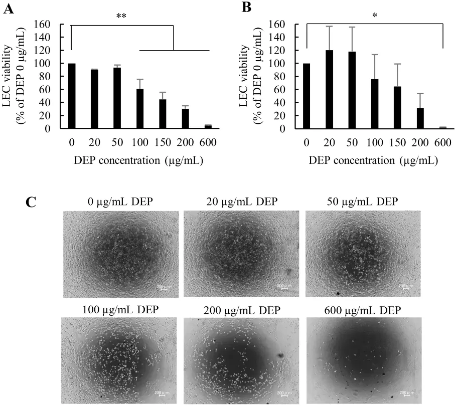
The cell viability of iLECs exposed to DEPs at 0 ~ 600 μg/mL was evaluated after either 6 or 24 h. As a result, cellular death appeared after 6 h. Moreover, exposure to > 100 μg/mL of DEPs induced significant cytotoxicity. At 600 μg/mL, almost all cells had died. A similar trend was also observed following 24 h of incubation (Fig. 1).
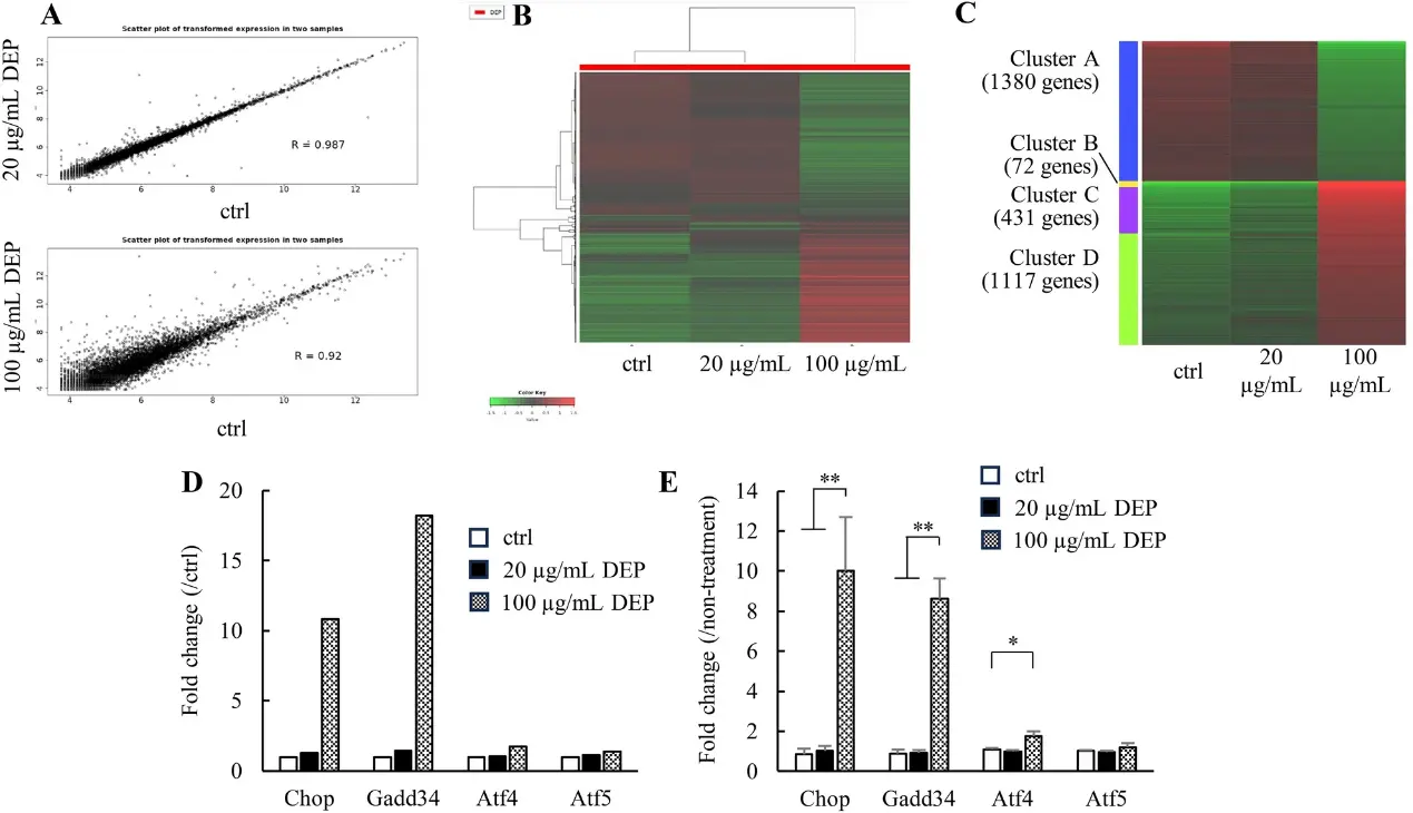
To determine what cascades are affected by DEP exposure to iLECs, we performed comprehensive RNA expression analysis. We identified 4 integrated stress response (ISR)-related genes that were increased in the transcriptome data: Chop (Ddit3, DNA-damage inducible transcript), Gadd34 (Ppr1r15a, protein phosphatase 1, regulatory subunit 15A), Atf4 (Creb2, activating transcription factor 4), and Atf5 (Atfa, activating transcription factor 5) (Fig. 2).
Ask a Question
Write your own review
Description: Immortalized Cynomolgus Monkey Pulmonary Artery Smooth Muscle Cells-GFP provided by Creative Bioarray have been developed by immortalizing cynomolgus monkey pulmonary artery smooth muscle cells with ...
Description: Immortalized Monkey Spleen Fibroblasts-SV40T were developed from monkey tissues transduced with a lentiviral expression vector containing the SV40T gene. The cell line was continuously cultured for ...
Description: Immortalized Monkey Bronchial Fibroblasts-GFP provided by Creative Bioarray have been developed by immortalizing monkey bronchial fibroblasts with SV40 Large T antigen and transfecting with tGFP. The ...
Description: Immortalized Canine Kidney Fibroblasts-SV40 have been obtained immortalizing Canine Kidney Fibroblasts with Lenti-SV40 Lentivirus. Immortalized cells were controlled passaging side by side with the ...
Description: Immortalized Monkey Primary Aortic Endothelial Cells-GFP provided by Creative Bioarray have been developed by immortalizing primary monkey aortic endothelial cells with SV40 Large T antigen and ...
Description: Immortalized Monkey Pancreatic Epithelial Cells-SV40T were developed from monkey tissues transduced with a lentiviral expression vector containing the SV40T gene. The cell line was continuously ...
- Adipose Tissue-Derived Stem Cells
- Human Neurons
- Mouse Probe
- Whole Chromosome Painting Probes
- Hepatic Cells
- Renal Cells
- In Vitro ADME Kits
- Tissue Microarray
- Tissue Blocks
- Tissue Sections
- FFPE Cell Pellet
- Probe
- Centromere Probes
- Telomere Probes
- Satellite Enumeration Probes
- Subtelomere Specific Probes
- Bacterial Probes
- ISH/FISH Probes
- Exosome Isolation Kit
- Human Adult Stem Cells
- Mouse Stem Cells
- iPSCs
- Mouse Embryonic Stem Cells
- iPSC Differentiation Kits
- Mesenchymal Stem Cells
- Immortalized Human Cells
- Immortalized Murine Cells
- Cell Immortalization Kit
- Adipose Cells
- Cardiac Cells
- Dermal Cells
- Epidermal Cells
- Peripheral Blood Mononuclear Cells
- Umbilical Cord Cells
- Monkey Primary Cells
- Mouse Primary Cells
- Breast Tumor Cells
- Colorectal Tumor Cells
- Esophageal Tumor Cells
- Lung Tumor Cells
- Leukemia/Lymphoma/Myeloma Cells
- Ovarian Tumor Cells
- Pancreatic Tumor Cells
- Mouse Tumor Cells