- Specification
- Background
- Scientific Data
- Q & A
- Customer Review
"Bovine Lung Cells, fetal" means lung cells isolated from the lungs of fetal cattle. In the airway and alveolar regions of the lung, fetal bovine lung cells mainly include alveolar cells and other cell types in the lungs. The morphology and structure of fetal bovine lung cells change significantly at different developmental stages. At an early stage, the structure is simple and the lung tissue is dominated by trachea and alveolar buds. At a later stage, the alveolar structure gradually appears, the alveolar wall becomes thinner, the alveolar cavity becomes larger, and the alveolar structure becomes more complex. In addition, fetal bovine lung cells also include a variety of cell types, including epithelial cells, fibroblasts, and immune cells such as macrophages and lymphocytes. Epithelial cells in alveoli (type I and type II alveolar cells) are involved in gas exchange and help maintain the exchange and regulation of gases in the alveoli. Type II alveolar cells secrete surfactant, which regulates alveolar surface tension and prevents alveolar collapse. Immune cells in the lungs (macrophages, lymphocytes, etc.) are involved in defending against pathogen invasion and inflammation. Fibroblasts and connective tissue cells in the lungs provide structural support and repair functions for the lungs. Studying the development and function of fetal bovine lung cells helps to understand the mechanisms of lung development, lung diseases, and lung repair.
M. bovis Infection Increased the ANXA2 Expression in the EBL Cells
Mycoplasma bovis (M. bovis), one of the most important pathogens causing bovine respiratory disease, colonizes the lower respiratory tracts and is responsible for severe pneumonia. However, the molecular pathogenesis of M. bovis is not well understood. Annexin A2 (ANXA2) is a calcium-dependent phospholipid-binding protein. Zhang's group investigated whether ANXA2 was involved in M. bovis adhesion, invasion, and inflammatory responses.
The invasion efficiency of M. bovis to EBL cells increased with increased multiplicity of infection (MOI) (1: 10 to 1:1000) and infection time (4 to 24 hpi) (Fig. 1A). The ANXA2 expression was significantly upregulated by M. bovis infection at both mRNA and protein levels, as demonstrated by quantitative polymerase chain reaction (qPCR) and western blotting. There was a gradual increase in ANXA2 mRNA expression during infection with time-dependent differences becoming statistically significant between 4 hours post-infection and 24 hours post-infection (Fig. 1B). The band density of ANXA2 protein was also upregulated with infection time (Fig. 1C, D). This indicates that ANXA2 expression was induced by M. bovis infection. In addition, the interacting protein of ANXA2, S100A10, was also significantly upregulated at both high MOI (MOI = 1: 100) and low MOI (MOI = 1:10) in infected cells, but not in the mock group.
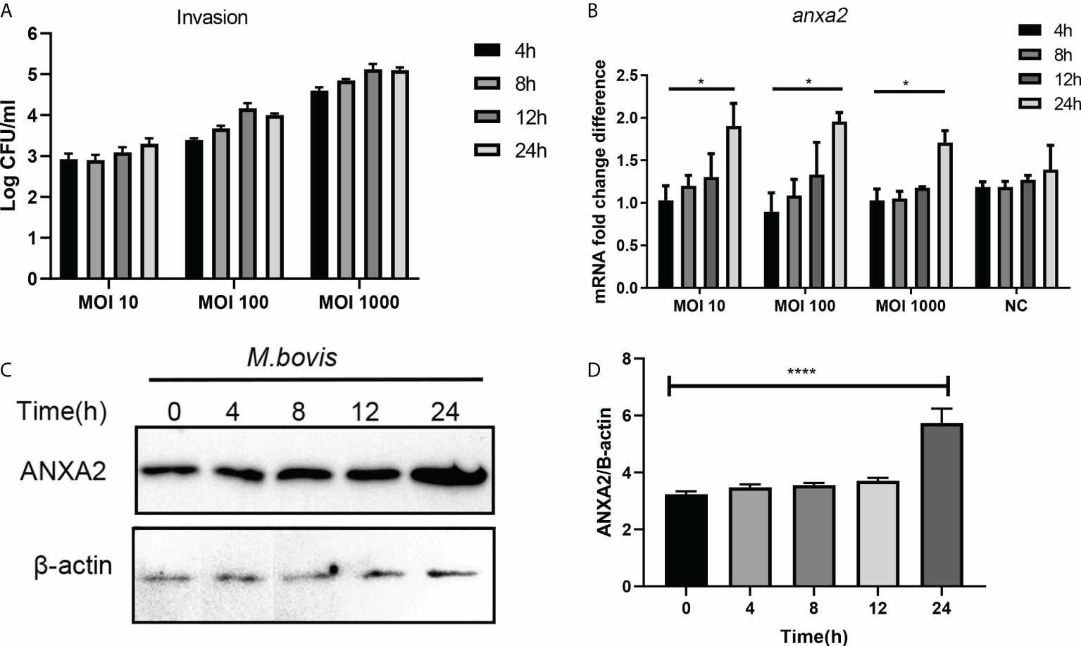 Fig. 1. M. bovis infection increased the ANXA2 expression in EBL cells (Zhang H, Lu D K, et al., 2022).
Characterization of EVs Derived from EBL Cells Infected with M. bovis
Mycoplasma bovis (M. bovis) mainly causes pneumonia, mastitis, and arthritis in cattle, resulting in enormous economic losses. Inflammation-related pathological damage is crucial to the pathogenesis of M. bovis. He's team focused on uncovering the role of extracellular vesicles (EVs) during M. bovis-host interaction while identifying the critical miRNAs within EVs.
EVs secreted from infected cells were purified without any cell debris to investigate the role of EVs in the regulation of host inflammatory response. The LDH assay kit was used to detect LDH released from EBL cells infected with M. bovis for 24 h at various MOIs (10, 20, 30, 50, 100, 200). LDH assay showed no significant cytotoxicity of M. bovis at MOIs 10 and 20 in EBL cells after 24 h (Fig. 2A). At MOI 20, cytotoxicity was unchanged at different infection times (Fig. 2B), and MOI 20 was chosen for the extraction of EVs. M. bovis survived in MEM and RPMI 1640 with 10% FBS at most 24 h. Transmission electron microscopy (TEM), dynamic light scattering (DLS), and western blotting revealed that EVs+ (infected) and EVs– (control) displayed cup-shaped double-membrane structures (Fig. 2C-D) with mean diameters of 276.84 nm and 190.64 nm, respectively (Fig. 2E-F), expressed EV markers CD63 and TSG101, but not the control protein calnexin (Fig. 2G).
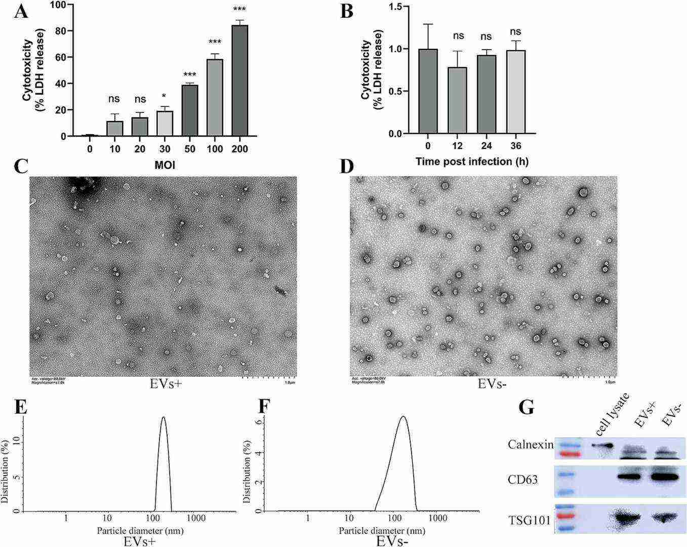 Fig. 2. Cytotoxicity of M. bovis PG45 against EBL cells and identification of EVs derived from EBL cells infected with M. bovis (He J X, Guo Y S, et al., 2025).
Ask a Question
Write your own review
Description: Bovine Preadipocytes from Creative Bioarray are isolated from the bovine adipose tissue. The method we use to isolate bovine preadipocytes were developed based on a combination of established and our ...

Description: Bovine Small Intestinal Epithelial Cells from Creative Bioarray are isolated from the bovine small intestinal tissue. The method we use to isolate bovine small intestinal epithelial cells were ...
Description: The BCHON cultures are on the base of bovine tissues. The BCHON are isolated according to known procedures. Then primary cultures are produced. After further culturing, the BCHON are frozen in a ...
Description: RFP Expressing Bovine Aortic Endothelial Cells (RFP-BAoECs) provided by Creative Bioarray are puromycin-selected from primary bovine aortic endothelial cells infected with lentivirus expressing RFP. ...

Description: Bovine Rumen Epithelial Cells from Creative Bioarray are isolated from the bovine rumen tissue. The method we use to isolate bovine rumen epithelial cells were developed based on a combination of ...
Description: Bovine Hepatic Stellate Cells from Creative Bioarray are isolated from the bovine liver tissue. The method we use to isolate bovine hepatic stellate cells were developed based on a combination of ...