
SD-1
Cat.No.: CSC-C1386
Species: Homo sapiens (Human)
Source: Blood; Peripheral Blood
Morphology: ovoid to round cells growing in suspension, singly or in clumps
Culture Properties: suspension
- Specification
- Background
- Scientific Data
- Q & A
- Customer Review
Immunology: CD3 -, CD10 -, CD13 -, CD19 +, CD20 +, CD37 +, cyCD79a +, CD80 +, HLA-DR +, sm/cyIgG +, sm/cyIgM -, sm/cykappa +, sm/cylambda -
Viruses: ELISA: reverse transc
SD-1 is a human B-cell precursor acute lymphoblastic leukemia (B-ALL) cell line that was originally derived from bone marrow of a pediatric patient with acute lymphoblastic leukemia. It is commonly used as an in vitro model system for molecular and cellular studies of B-lineage leukemia, in particular, aggressive and/or therapy-resistant forms of the disease. SD-1 harbors chromosomal and genetic changes typically associated with high-risk ALL, including the Philadelphia chromosome, t(9;22)(q34;q11), and expression of the BCR-ABL fusion oncogene. Consequently, SD-1 has constitutive activation of tyrosine kinase signaling cascades associated with cell proliferation, survival, and anti-apoptotic mechanisms. The cell line is positive for B-cell marker expression including CD19 and CD10, consistent with the B-cell precursor immunophenotype.
Functionally, SD-1 is responsive to tyrosine kinase inhibitors and to standard chemotherapeutic agents. For these reasons, the cell line is a useful tool for studies of drug sensitivity, resistance, and signaling pathway dependencies in Ph-positive ALL. SD-1 is also commonly used for preclinical drug screening of novel targeted agents and novel combination treatment approaches, as well as for studies of leukemogenesis.
The Influence of the Cultivation Conditions on the Proliferation and Metabolism of the Leukemia Cells
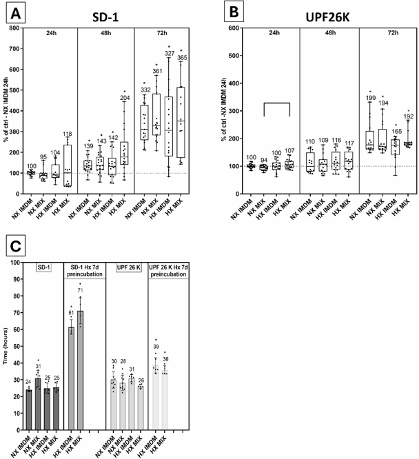
Leukemia is influenced by the hypoxic bone marrow microenvironment, affecting disease progression and treatment resistance. Sikorová et al. investigated the interactions between leukemic cell lines (SD-1 and UPF26K) and feeder cells (NHDF and hMSCs) under normoxic and hypoxic conditions to understand their impact on cell proliferation and metabolism.
The proliferation of SD-1 and UPF26K cells was tested in two media-IMDM and αMEM + IMDM (1:1)-under NX (20% O2) and HX (1% O2) conditions. SD-1 cells (Fig. 1A) showed similar growth in all media and oxygen conditions during short-term (24-72 h) cultivation, with a doubling time of about 25 hours (Fig. 1C). Pre-incubating SD-1 cells under HX for 7 days tripled their doubling time to about 70 hours. UPF26K cells (Fig. 1B) also showed similar proliferation trends and doubling times (about 28 hours) in all conditions (Fig. 1C). Pre-incubating UPF26K cells under HX for 7 days reduced their proliferation, but less than SD-1 cells, with a doubling time of about 38 hours.
Ask a Question
Write your own review
- You May Also Need

Description: Established in 2007 from the bone marrow mononuclear cells of an 82-year-old Japanese man with diffuse large B-cell lymphoma in the leukemic phase

Description: Established from the bone marrow of a 28-year-old man who developed the terminal leukemic phase of lymphosarcoma in 1976

Description: This cell line was derived from the bone marrow aspirate of a 59 year old male with erythroleukemia that became acute myelogenous leukaemia.The cells form colonies in soft-agar in the presence of ...

Description: Established from the pleural effusion of a 24-year-old woman with recurrent anaplastic large cell lymphoma (ALCL); cells were described to clonally derive from T-lineage lymphoid cells and to be ...

Description: Established from a 37-year-old man at second (refractory/terminal) relapse of Hodgkin lymphoma (nodular sclerosing -> lymphocyte depleted/stage IIISA -> stage IV) after both combined chemo- and ...

Description: Established from the peripheral blood of a 10-year-old Caucasian boy with acute lymphoblastic leukemia (pre B-ALL) at diagnosis in 1993
- Adipose Tissue-Derived Stem Cells
- Human Neurons
- Mouse Probe
- Whole Chromosome Painting Probes
- Hepatic Cells
- Renal Cells
- In Vitro ADME Kits
- Tissue Microarray
- Tissue Blocks
- Tissue Sections
- FFPE Cell Pellet
- Probe
- Centromere Probes
- Telomere Probes
- Satellite Enumeration Probes
- Subtelomere Specific Probes
- Bacterial Probes
- ISH/FISH Probes
- Exosome Isolation Kit
- Human Adult Stem Cells
- Mouse Stem Cells
- iPSCs
- Mouse Embryonic Stem Cells
- iPSC Differentiation Kits
- Mesenchymal Stem Cells
- Immortalized Human Cells
- Immortalized Murine Cells
- Cell Immortalization Kit
- Adipose Cells
- Cardiac Cells
- Dermal Cells
- Epidermal Cells
- Peripheral Blood Mononuclear Cells
- Umbilical Cord Cells
- Monkey Primary Cells
- Mouse Primary Cells
- Breast Tumor Cells
- Colorectal Tumor Cells
- Esophageal Tumor Cells
- Lung Tumor Cells
- Leukemia/Lymphoma/Myeloma Cells
- Ovarian Tumor Cells
- Pancreatic Tumor Cells
- Mouse Tumor Cells