PLC/PRF5
Cat.No.: CSC-C9581L
Species: Homo sapiens (Human)
Source: Liver
Morphology: epithelial-like
Culture Properties: monolayer
- Specification
- Background
- Scientific Data
- Q & A
- Customer Review
vWA: 15,16
FGA: 19.2,25
Amelogenin: X
TH01: 7,8
TPOX: 8
CSF1P0: 10
D5S818: 12
D13S317: 11,12
D7S820: 9,11
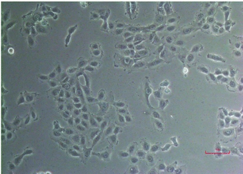
PLC/PRF/5 (AKA Alexander cells) is a human hepatocellular carcinoma (HCC) cell line that was originally established from the primary liver tumor of a 24-year-old man. It was one of the first human hepatocellular carcinoma cell lines shown to express hepatitis B virus (HBV) surface antigen (HBsAg), consistent with chronic HBV infection as an etiologic factor in human liver cancer. PLC/PRF/5 cells do not produce infectious viral particles but have integrated HBV DNA sequences that drive stable expression of HBsAg.
These cells have an epithelial-like, polygonal morphology and are adherent monolayer cells with distinct cell-cell borders. PLC/PRF/5 cells grow rapidly under standard tissue culture conditions, are typically maintained in MEM or DMEM media supplemented with fetal bovine serum, and maintain many hepatocyte-associated characteristics including expression of liver-specific proteins and metabolic enzymes. However, PLC/PRF/5 cells, like many tumor cells, are genetically unstable. At the molecular level, these cells harbor chromosomal abnormalities and mutations relevant to hepatocarcinogenesis, including alterations in pathways regulating cell cycle control, apoptosis, and oncogenic signaling. Thus, the PLC/PRF/5 cell line is useful for studying virus-host interactions and virus-associated carcinogenesis, in addition to being a model system for the study of the role of viral factors in the development of liver tumors.
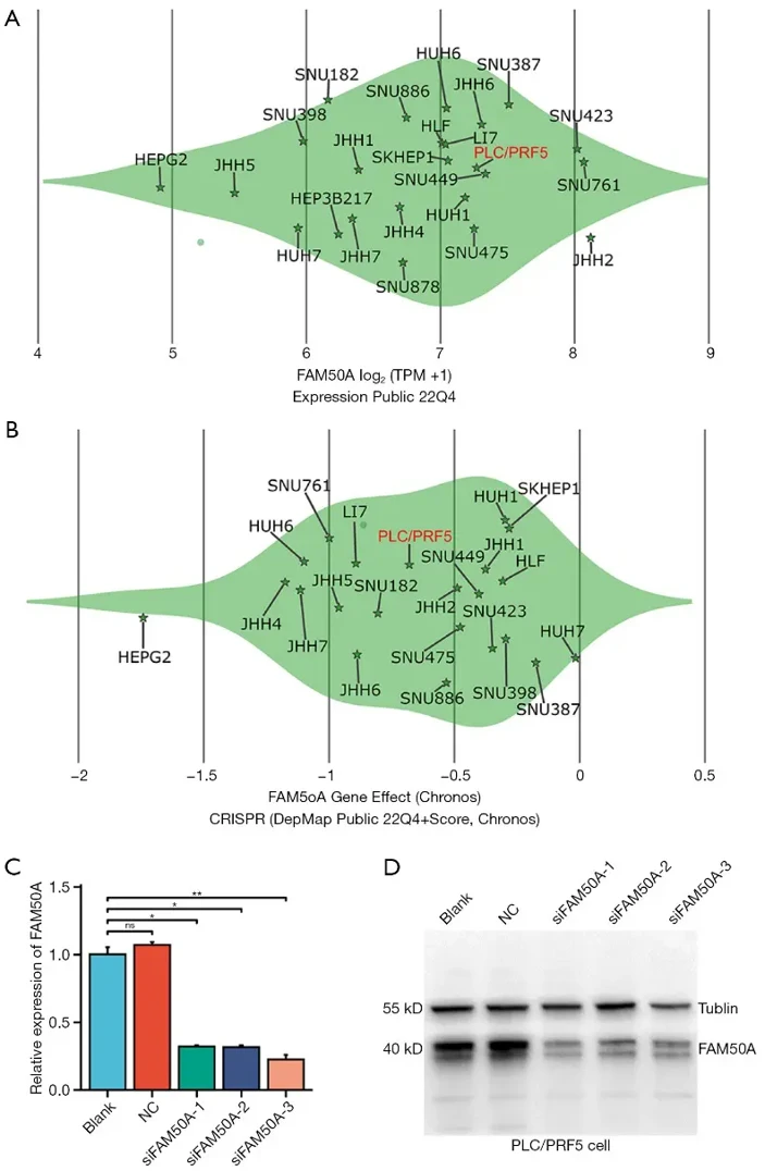
FAM50A Knockout Suppresses Growth and Survival in Hepatocarcinoma Cells
Hepatocellular carcinoma (HCC) is one of the most lethal malignancies with few effective therapies. FAM genes are candidate regulators of tumorigenesis, but their involvement in HCC is not well known. Here, Wu's team performed a comprehensive investigation on the expression profiles and functional roles of FAM genes in HCC.
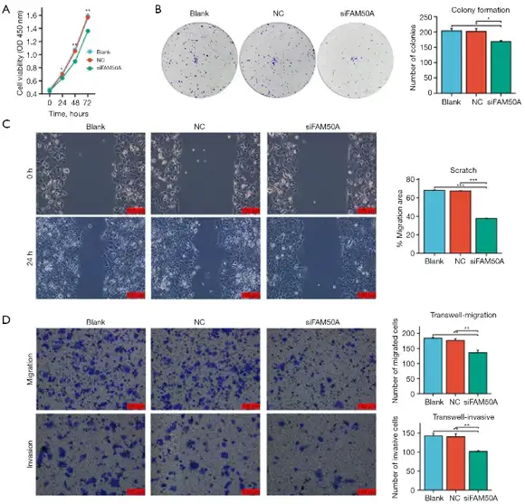
The FAM50A expression level in different human hepatocarcinoma cells varied from 4.91 in HEPG2 to 8.12 in JHH2 (Fig. 1A). Knocking out of FAM50A greatly decreased the probability of cell survival lower than -0.5 in 11 hepatocarcinoma cells (Fig. 1B), indicating the critical role of FAM50A for HCC cell growth and survival. PLC/PRF5 cells, with intermediate FAM50A expression (7.27) and RNAi effect (-0.71), were chosen for further study. PLC/PRF5 cells were transfected with three siFAM50A sequences, significantly reducing FAM50A expression at both mRNA and protein levels (Fig. 1C and D). siFAM50A-1 showed the strongest inhibitory effect and was selected for subsequent experiments. CCK-8 and colony formation assays revealed reduced viability and proliferation in FAM50A-knockdown PLC/PRF5 cells (Fig. 2A and B). Scratch and Trans-well migration assays indicated impaired migration (Fig. 2C), and the Trans-well invasion assay showed fewer invasive cells in the siFAM50A group compared to controls (Fig. 2D).
Ask a Question
Write your own review
- You May Also Need
Description: Ito (fat-storing) cells, stellate-shaped mesenchymal cells that exist in the space of Disse of the liver and contain many fat droplets in cytoplasm.

Description: This is one cell line out of a series of glioblastoma cell lines established by PD Dr. Michael Linnebacher.
Description: Human bile duct cell line established from ascites of the tumor patient who had differentiated adenocarcinoma.


Description: Human cell line derived from cholangiocellular carcinoma. Cell growth is slow.
- Adipose Tissue-Derived Stem Cells
- Human Neurons
- Mouse Probe
- Whole Chromosome Painting Probes
- Hepatic Cells
- Renal Cells
- In Vitro ADME Kits
- Tissue Microarray
- Tissue Blocks
- Tissue Sections
- FFPE Cell Pellet
- Probe
- Centromere Probes
- Telomere Probes
- Satellite Enumeration Probes
- Subtelomere Specific Probes
- Bacterial Probes
- ISH/FISH Probes
- Exosome Isolation Kit
- Human Adult Stem Cells
- Mouse Stem Cells
- iPSCs
- Mouse Embryonic Stem Cells
- iPSC Differentiation Kits
- Mesenchymal Stem Cells
- Immortalized Human Cells
- Immortalized Murine Cells
- Cell Immortalization Kit
- Adipose Cells
- Cardiac Cells
- Dermal Cells
- Epidermal Cells
- Peripheral Blood Mononuclear Cells
- Umbilical Cord Cells
- Monkey Primary Cells
- Mouse Primary Cells
- Breast Tumor Cells
- Colorectal Tumor Cells
- Esophageal Tumor Cells
- Lung Tumor Cells
- Leukemia/Lymphoma/Myeloma Cells
- Ovarian Tumor Cells
- Pancreatic Tumor Cells
- Mouse Tumor Cells