
MONO-MAC-1
Cat.No.: CSC-C0353
Species: Homo sapiens (Human)
Source: Blood; Peripheral Blood
Morphology: single, round/multiformed cells in suspension
Culture Properties: suspension
- Specification
- Background
- Scientific Data
- Q & A
- Customer Review
- Documents
Ship in dry ice; Store in liquid nitrogen
Immunology: CD3 -, CD13 +, CD14 -, CD15 +, CD19 -, CD34 -, cyCD68 +, HLA-DR +
Viruses: ELISA: reverse transcriptase negative; PCR: EBV -, HBV -, HCV -, HHV-8 -, HIV -, HTLV-I/II -, MLV -, SMRV -
MONO-MAC-1 cells are a cell line that was established from the peripheral blood of a 64-year-old man diagnosed with acute monocytic leukemia (AML FAB M5) at relapse in 1985. These cells were obtained following myeloid metaplasia, which refers to the abnormal transformation of bone marrow cells into cells of the myeloid lineage.
The MONO-MAC-1 cell line is a sister cell line of MONO-MAC-6 (DSM ACC 124), meaning they share a common origin or are derived from the same parental cell population. However, MONO-MAC-1 cells are described as being less differentiated than MONO-MAC-6 cells.
Establishing MONO-MAC-1 cells has provided researchers with a useful tool for investigating the molecular mechanisms underlying ALL and HIV-1 infection. These cells have been utilized in numerous studies aiming to uncover the factors involved in the differentiation of monocytes, the pathogenesis of HIV-1, and the development of novel therapeutic interventions for leukemia and HIV/AIDS.
Modulation of CCR7 Receptor Expression in Mono-MAC-1 Cells by LXRα Activation and PGE2
Cell migration via chemokine receptor CCR7 expression is an essential function of the immune system. Prostaglandin E2 (PGE2), an important immunomodulatory molecule, increases CCR7 expression and function in monocytes. MONO-MAC-1 (MM-1) is a human cell line with the properties of blood monocytes that can be used as a model system to study monocytic functions in vitro.
MM-1 cells were treated with 1 μM PGE2 and 1 μM T0901317, a synthetic liver X receptor α (LXRα) agonist, for 8 or 24 h. As previously observed, PGE2 induces CCR7 mRNA expression with a maximal effect after stimulation for 8 h. Treatment of MM-1 cells with T0901317 alone does not modify the expression of CCR7 mRNA (Fig. 1a). An increase in LXRα levels following treatment with PGE2 or T0901317 alone. mRNA production plateaued after 8 h of stimulation with PGE2 or T0901317 alone (Fig. 1b). ABCG1 mRNA levels were augmented when cells were treated with T0901317 alone. PGE2 did not affect ABCG1 transcription (Fig. 1c).
MM-1 cells were treated with the stimulants for 24 or 48 hours before migration through polycarbonate filters (5 μm pore size) for 4 h. The chemotaxis assay results showed that PGE2 increases MM-1 cell migration to both CCR7 natural ligands CCL19 (Fig. 2a) and CCL21 (Fig. 2b) after treatment for 24 h. The LXR agonist alone did not affect the migratory capacity of MM-1 cells in response to CCL19 whereas 24 h treatment with T0901317 significantly increased migration in response to CCL21. To further investigate the effects of PGE2 and T0901317 on CCR7 expression, migration assays were repeated with freshly isolated human blood monocytes. Results showed that monocyte migration toward CCL19 and CCL21 is increased following the treatment of blood monocytes with PGE2 and T0901317 (Fig. 3).
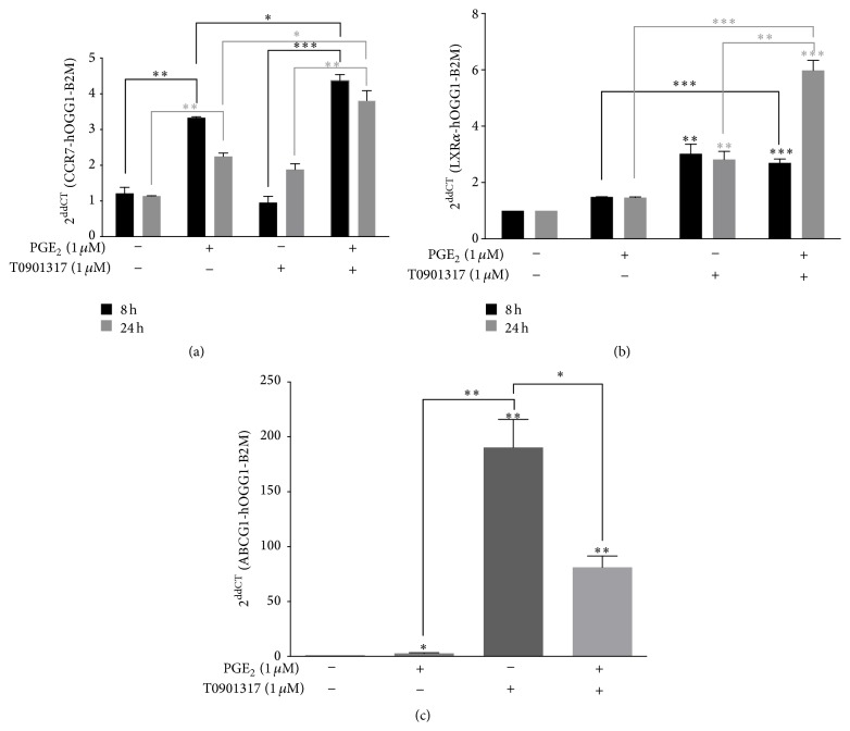 Fig. 1 Effects of PGE2 and T0901317 on CCR7, ABCG1, and LXRα transcription in MM-1 cells. (Tanné B, et al., 2015)
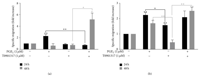 Fig. 2 PGE2 alone and in combination with T0901317 induces functional CCR7-specific migration of MM-1 cells. (Tanné B, et al., 2015)
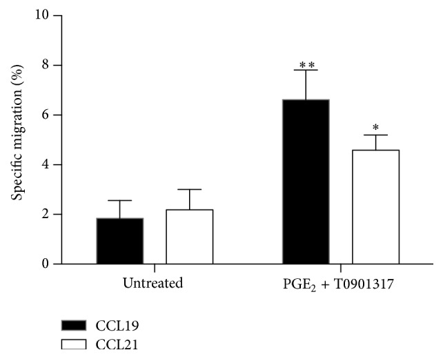 Fig. 3 Freshly isolated monocytes migrate toward the CCR7-specific ligands CCL19 and CCL21. (Tanné B, et al., 2015)
CSF-1R Upregulation with Tyrosine Kinase in Mono-Mac-1 Cell Lines
The oncogenic potential of colony-stimulating factor 1 receptor (CSF-1R) has been well described, while its relevance for human AML is still undetermined. To assess the relevance of CSF-1R in AML, three myeloid cell lines, Mono-Mac 1, THP-1, and U937, were treated with compounds that affect the CSF-1R and other pathways. After allowing the cells to grow for three days, only Mono-Mac 1 cells demonstrated reduced proliferation with CSF-1R inhibitor (cFMS-I) treatment. In Mono-Mac 1 cells, cFMS-I increased the G1 population from 57.92% to 64.04% (Fig. 4B), Sunitinib increased G1 cells from 57.92% to 63.4% (Fig. 4C), and U0126 increased the G1 population from 57.92% to 62.6% (Fig. 4D).
The effects of cFMS-I (1 μM), sunitinib (0.1 μM), and U0126 (10 μM) on ERK activity were compared in Mono-Mac 1 cells with and without CSF-1 treatment (Fig. 5B). As shown in Fig. 5A, the Mono-Mac 1cells had high ERK activity, and CSF-1 induced slight ERK activation. cFMS-I, sunitinib, and U0126 all inhibited ERK activity. sunitinib caused the greatest inhibition of ERK, followed by cFMS-I, and U0126 had the smallest effect on ERK. CSF-1R was detected in Mono-Mac 1 cells (Fig. 5C). The CSF-1R was down-regulated after 5 minutes of treatment with CSF-1 (Fig. 5D). Mono-Mac 1 cells were treated with cFMS-I (0.01-1 μM) and sunitinib (0.01-1μM) (Fig. 5E). By western blot, both treatments did indeed show a profound and dose-dependent increase in the amount of total CSF-1R receptor in Mono-Mac 1 cells (Fig. 5F).
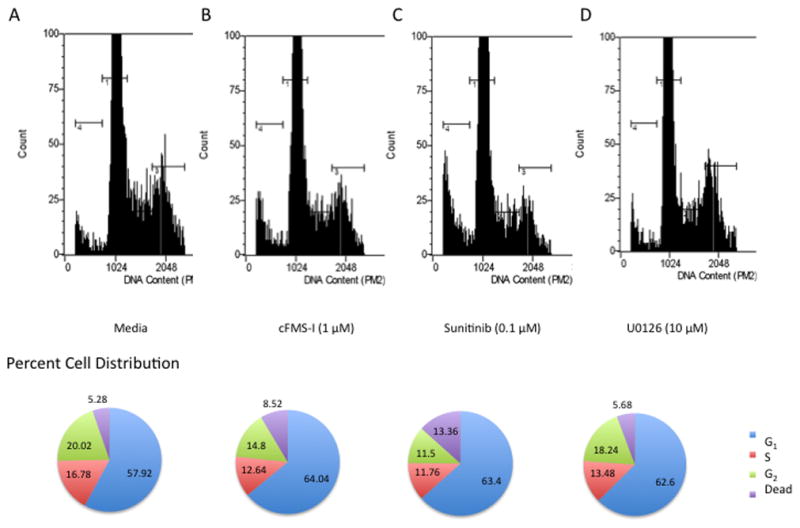 Fig. 4 The effect of colony-stimulating factor 1 receptor inhibitor, sunitinib, and U0126 on Mono-Mac 1 cell cycle distribution. (Kogan M, et al., 2012)
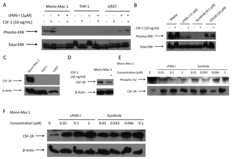 Fig. 5 Cell signaling and colony-stimulating factor 1 receptor (CSF-1R) receptor function with drug treatment in acute myelogenous leukemia cell lines. (Kogan M, et al., 2012)
Ask a Question
Write your own review
- You May Also Need

Description: Established in 2007 from the bone marrow mononuclear cells of an 82-year-old Japanese man with diffuse large B-cell lymphoma in the leukemic phase

Description: Established from the bone marrow of a 28-year-old man who developed the terminal leukemic phase of lymphosarcoma in 1976

Description: This cell line was derived from the bone marrow aspirate of a 59 year old male with erythroleukemia that became acute myelogenous leukaemia.The cells form colonies in soft-agar in the presence of ...

Description: Established from the pleural effusion of a 24-year-old woman with recurrent anaplastic large cell lymphoma (ALCL); cells were described to clonally derive from T-lineage lymphoid cells and to be ...

Description: Established from a 37-year-old man at second (refractory/terminal) relapse of Hodgkin lymphoma (nodular sclerosing -> lymphocyte depleted/stage IIISA -> stage IV) after both combined chemo- and ...

Description: Established from the peripheral blood of a 10-year-old Caucasian boy with acute lymphoblastic leukemia (pre B-ALL) at diagnosis in 1993
- Adipose Tissue-Derived Stem Cells
- Human Neurons
- Mouse Probe
- Whole Chromosome Painting Probes
- Hepatic Cells
- Renal Cells
- In Vitro ADME Kits
- Tissue Microarray
- Tissue Blocks
- Tissue Sections
- FFPE Cell Pellet
- Probe
- Centromere Probes
- Telomere Probes
- Satellite Enumeration Probes
- Subtelomere Specific Probes
- Bacterial Probes
- ISH/FISH Probes
- Exosome Isolation Kit
- Human Adult Stem Cells
- Mouse Stem Cells
- iPSCs
- Mouse Embryonic Stem Cells
- iPSC Differentiation Kits
- Mesenchymal Stem Cells
- Immortalized Human Cells
- Immortalized Murine Cells
- Cell Immortalization Kit
- Adipose Cells
- Cardiac Cells
- Dermal Cells
- Epidermal Cells
- Peripheral Blood Mononuclear Cells
- Umbilical Cord Cells
- Monkey Primary Cells
- Mouse Primary Cells
- Breast Tumor Cells
- Colorectal Tumor Cells
- Esophageal Tumor Cells
- Lung Tumor Cells
- Leukemia/Lymphoma/Myeloma Cells
- Ovarian Tumor Cells
- Pancreatic Tumor Cells
- Mouse Tumor Cells