LP-1
Cat.No.: CSC-C6221X
Species: Homo sapiens (Human)
Source: Blood; Peripheral Blood
Morphology: single, elongated, snake-like cells in suspension
Culture Properties: suspension
- Specification
- Background
- Scientific Data
- Q & A
- Customer Review
Immunology: CD3 -, CD10 -, CD13 -, CD19 -, CD20 -, CD34 -, CD37 -, CD38 +, cyCD79a -, CD80 -, CD138 +, HLA-DR +
LP-1 is a human multiple myeloma cell line that was derived from the peripheral blood of a patient with plasma cell leukemia, a very aggressive subtype of multiple myeloma. LP-1 is a malignant plasma cell-derived cell line frequently used for mechanistic in vitro studies of myeloma progression, plasma cell biology, and response to therapy.
LP-1 cells grow as a suspension culture and phenotypically resemble plasma cells, including expression of CD38, CD138 (syndecan-1), and light chains of immunoglobulins. The cell line has stable proliferation and harbors molecular and phenotypic characteristics that are emblematic of advanced disease. LP-1 cells are typically cultured in RPMI-1640 media supplemented with fetal bovine serum and standard cell culture growth factors.
LP-1 cells have been used in research on hematological malignancies and are frequently utilized for mechanistic studies of oncogenic signaling, regulation of apoptosis, and drug resistance in myeloma. These cells are also often used to screen for novel antimyeloma agents, for studies involving proteasome inhibition and myeloma interactions with bone marrow stroma. Because of their reproducibility, clinical relevance, and defined plasma cell phenotype, LP-1 cells are a useful model for basic, translational, and preclinical studies of multiple myeloma.
Functional Effects of TBC1 Domain Containing Kinase Deletion in Immortalized B Cells and Plasma Cells
TBC1 domain containing kinase (TBCK) is highly expressed in neurons and glial cells, and its mutations cause intellectual disability and hypotonia. TBCK is part of the FERRY complex involved in mRNA transport and mTORC1 signaling. Here, Beck et al. generated TBCK knockout cell lines (Raji and LP-1 cells) to study its impact on B cells and plasma cells.
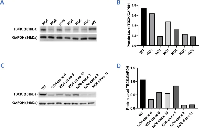
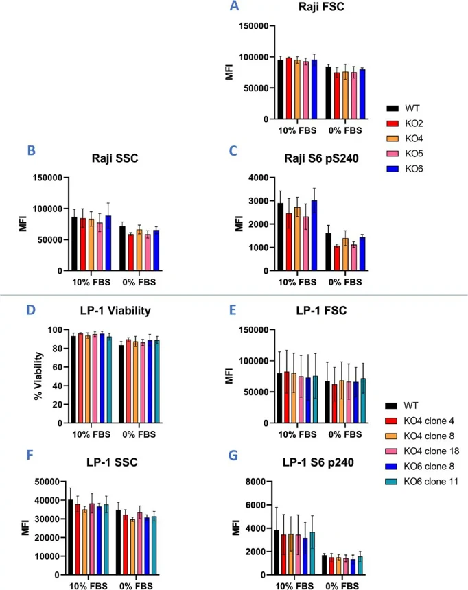
In Raji cells, lentiviral transduction with guide RNAs KO2, KO4, KO5, or KO6 significantly reduced TBCK expression (Fig. 1A-B). In LP-1 cells, initial TBCK knockdown was modest, with only KO4 and KO6 showing some reduction. To improve knockdown in LP-1 cells, Beck et al. generated single-cell clones, and some of these clones had satisfactory TBCK knockdown (Fig. 1C-D). They then used flow cytometry to see if the effects of TBCK knockdown seen in other cell models could be reproduced in Raji or LP-1 cells. They grew these cells for 24 hours in media with either 10% FBS or 0% FBS to mimic conditions with and without EGFR signaling. Surprisingly, neither Raji nor LP-1 cells showed any sensitivity to TBCK loss under normal or serum-free conditions. All modified cell lines had similar viability, cell size, and S6 phosphorylation levels. They also had similar side-scatter profiles, indicating no increase in cellular granularity or autophagosome accumulation in TBCK knockout cells. As expected, both cell lines had reduced S6 phosphorylation after serum withdrawal, especially in LP-1 cells, suggesting a greater reliance on EGF for homeostasis.
Ask a Question
Write your own review
- You May Also Need

Description: Established in 2007 from the bone marrow mononuclear cells of an 82-year-old Japanese man with diffuse large B-cell lymphoma in the leukemic phase

Description: Established from the bone marrow of a 28-year-old man who developed the terminal leukemic phase of lymphosarcoma in 1976

Description: This cell line was derived from the bone marrow aspirate of a 59 year old male with erythroleukemia that became acute myelogenous leukaemia.The cells form colonies in soft-agar in the presence of ...

Description: Established from the pleural effusion of a 24-year-old woman with recurrent anaplastic large cell lymphoma (ALCL); cells were described to clonally derive from T-lineage lymphoid cells and to be ...

Description: Established from a 37-year-old man at second (refractory/terminal) relapse of Hodgkin lymphoma (nodular sclerosing -> lymphocyte depleted/stage IIISA -> stage IV) after both combined chemo- and ...

Description: Established from the peripheral blood of a 10-year-old Caucasian boy with acute lymphoblastic leukemia (pre B-ALL) at diagnosis in 1993
- Adipose Tissue-Derived Stem Cells
- Human Neurons
- Mouse Probe
- Whole Chromosome Painting Probes
- Hepatic Cells
- Renal Cells
- In Vitro ADME Kits
- Tissue Microarray
- Tissue Blocks
- Tissue Sections
- FFPE Cell Pellet
- Probe
- Centromere Probes
- Telomere Probes
- Satellite Enumeration Probes
- Subtelomere Specific Probes
- Bacterial Probes
- ISH/FISH Probes
- Exosome Isolation Kit
- Human Adult Stem Cells
- Mouse Stem Cells
- iPSCs
- Mouse Embryonic Stem Cells
- iPSC Differentiation Kits
- Mesenchymal Stem Cells
- Immortalized Human Cells
- Immortalized Murine Cells
- Cell Immortalization Kit
- Adipose Cells
- Cardiac Cells
- Dermal Cells
- Epidermal Cells
- Peripheral Blood Mononuclear Cells
- Umbilical Cord Cells
- Monkey Primary Cells
- Mouse Primary Cells
- Breast Tumor Cells
- Colorectal Tumor Cells
- Esophageal Tumor Cells
- Lung Tumor Cells
- Leukemia/Lymphoma/Myeloma Cells
- Ovarian Tumor Cells
- Pancreatic Tumor Cells
- Mouse Tumor Cells