
F-36P
Cat.No.: CSC-C0551
Species: Homo sapiens (Human)
Source: Pleural Effusion
Morphology: polymorph cells growing as adherent cells and singly in suspension
- Specification
- Background
- Scientific Data
- Q & A
- Customer Review
Immunology: CD3 -, CD13 +, CD14 -, CD15 -, CD19 -, CD33 +, CD34 +, CD41 -, CD42 -, CD71 +, CD235a +
Viruses: PCR: EBV -, HBV -, HCV -, HIV -, HTLV-I/II -, SMRV -
F-36P is a cell line of human leukemia. It was isolated from the bone marrow of a 68-year-old man with acute myeloid leukemia (AML M6) and myelodysplastic syndrome (MDS) (subtype: refractory anemia with hyperleukocytosis) when he was diagnosed. And it was established in 1989 from the pleural effusion of this patient. The F-36P cell line has polymorphic morphology and can be cultured in adherent and suspended forms. F-36P cells express multiple blood development markers CD34, CD13, and CD33 while lacking T cell markers B cell markers and mature myeloid monocyte markers. These features suggest that F-36P has a certain degree of multipotency, but is not representative of T or B cells.
As a red-white cell line, the main application of F-36P is to model the abnormal differentiation process of erythroid progenitor cells in human bone marrow. Under normal physiological conditions, erythroid progenitor cells are induced to differentiate into mature red blood cells in a process that responds to some signals, such as erythropoietin (EPO). However, in the F-36P cell line, the induction of cell differentiation is IL-3-dependent. On the other hand, in some circumstances (such as the application of the active ingredient of realgar, As₂S₃), it can also cause apoptosis and differentiation. In addition, F-36P cells are also used to model megakaryocyte differentiation induced by thrombopoietin (TPO) and hematopoietic cell differentiation mediated by Ras signaling pathway.
Levels of DNMTs and Methyl-Binding Proteins Are Higher in F-36P/AZA Cells than in F-36P Cells
MDS is an age-related clonal disease with poor prognosis. Azacitidine (AZA) is a standard of care for MDS but only 30–40 % of patients respond to the treatment and mechanisms of drug resistance are still unknown. Kim et al. attempted to identify DNA-methylation-based biomarkers to predict AZA resistance, with a view to implementing an earlier treatment modification.
In order to identify the mechanisms of AZA resistance, they established an AZA-resistant cell model using F-36P cell line. As shown in Fig. 1, F-36P/AZA cells were highly resistant to AZA when compared to F-36P cells. The IC50 of AZA for F-36P/AZA was 125 μmol/L, which was 125 times higher than that of F-36P (1 μmol/L) (Fig. 1D). In a previous report, up-regulated mRNA levels of DNMT1, DNMT3a, and DNMT3b were found to be associated with AZA resistance of leukemia cells. They also found that the expression of DNMTs was higher in F-36P/AZA cells than in F-36P cells (Fig. 2A and B). The expression of DNMT3B was significantly higher than that of DNMT1 and DNMT3A. In addition, DNMTs, MBD2, and MeCP2 proteins were also more highly expressed in F-36P/AZA cells (Fig. 2C–H). Therefore, they confirmed that the expression of DNMTs was significantly upregulated in F-36P/AZA cells.
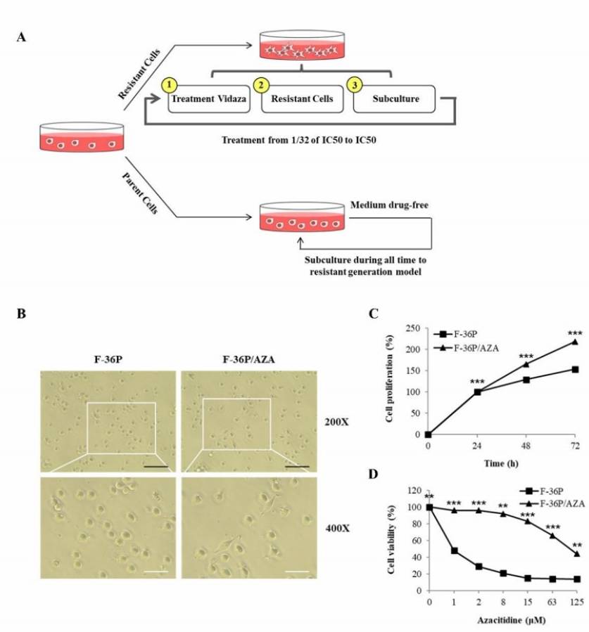 Fig. 1. Establishment of azacitidine resistant cell line (Kim DY, Shin D-Y, et al., 2024).
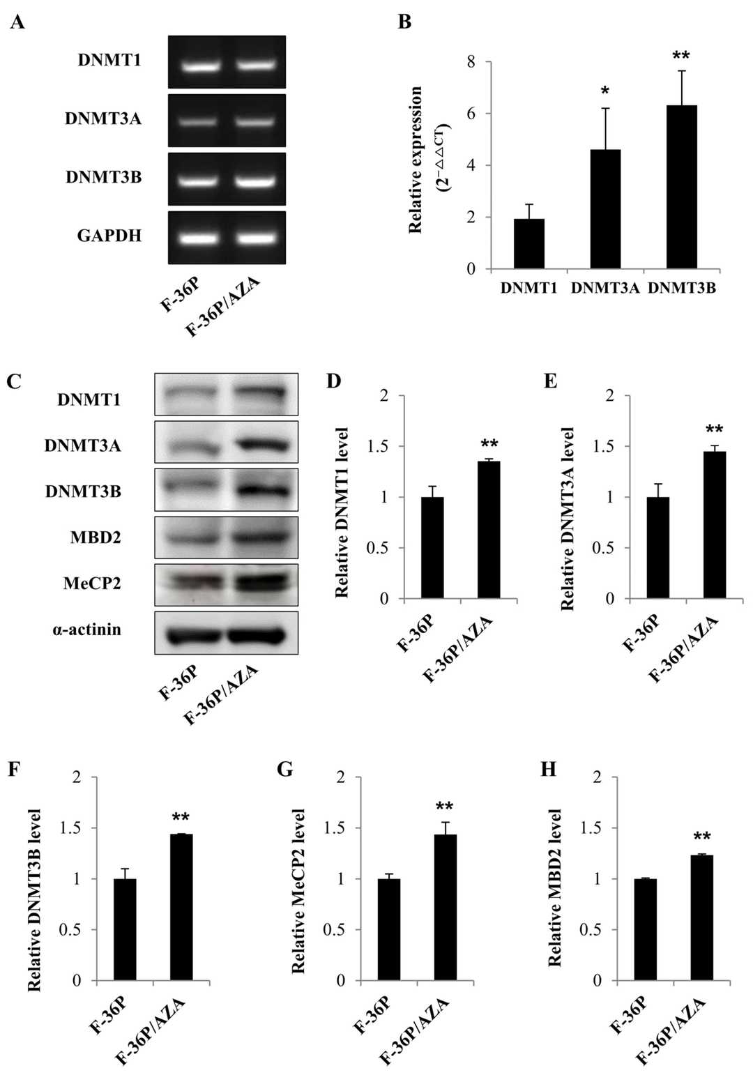 Fig. 2. Gene expression and protein levels of DNMTs in F-36P and F-36P/AZA cells (Kim DY, Shin D-Y, et al., 2024).
Gene Expression Profiles Identify Biomarkers of Resistance to Decitabine in Myelodysplastic Syndromes
MDS is an incurable clonal stem-cell disease. DEC is only useful for a part of patients and the development of resistance is related to the short survival. Kim et al. attempted to identify gene-expression signatures that will predict resistance to DEC and lead to a more personalized therapy.
To analyze the mechanism of DEC resistance, they established a DEC-resistant cell line (F-36P/DEC) from the F-36P cell line (Fig. 3A). F-36P cells were round and were seen as single cells under the light microscope, but F-36P/DEC cells aggregated with each other (Fig. 3B). F-36P/DEC cells were able to maintain stable proliferation for 72 h (Fig. 3C). The IC50 for DEC was 17.27 μmol/L in F-36P cells but exceeded 30 μmol/L in F-36P/DEC cells; accurate IC50 calculation wasn't possible even at 1000 μmol/L (Fig. 3D). F-36P/DEC cells had a higher proportion of cells in the G2/M phase than F-36P cells (Fig. 3E). Taken together, the results show that the F-36P/DEC model was greatly resistant to DEC and had different properties from F-36P cells. To find genes associated with DEC resistance, NanoString analysis was performed to identify DEGs between F-36P and F-36P/DEC, and 189 DEGs were found (Fig. 4A and B). The heatmap (Fig. 4A) and volcano plot (Fig. 4B) show this difference, and a scatter plot revealed more deviation from linearity with larger expression differences (Fig. 4C). GO terms found for DEGs were mainly related to transcription regulation, cell cycle, proliferation, nucleus, extracellular region, transcription-factor binding and kinase activity.
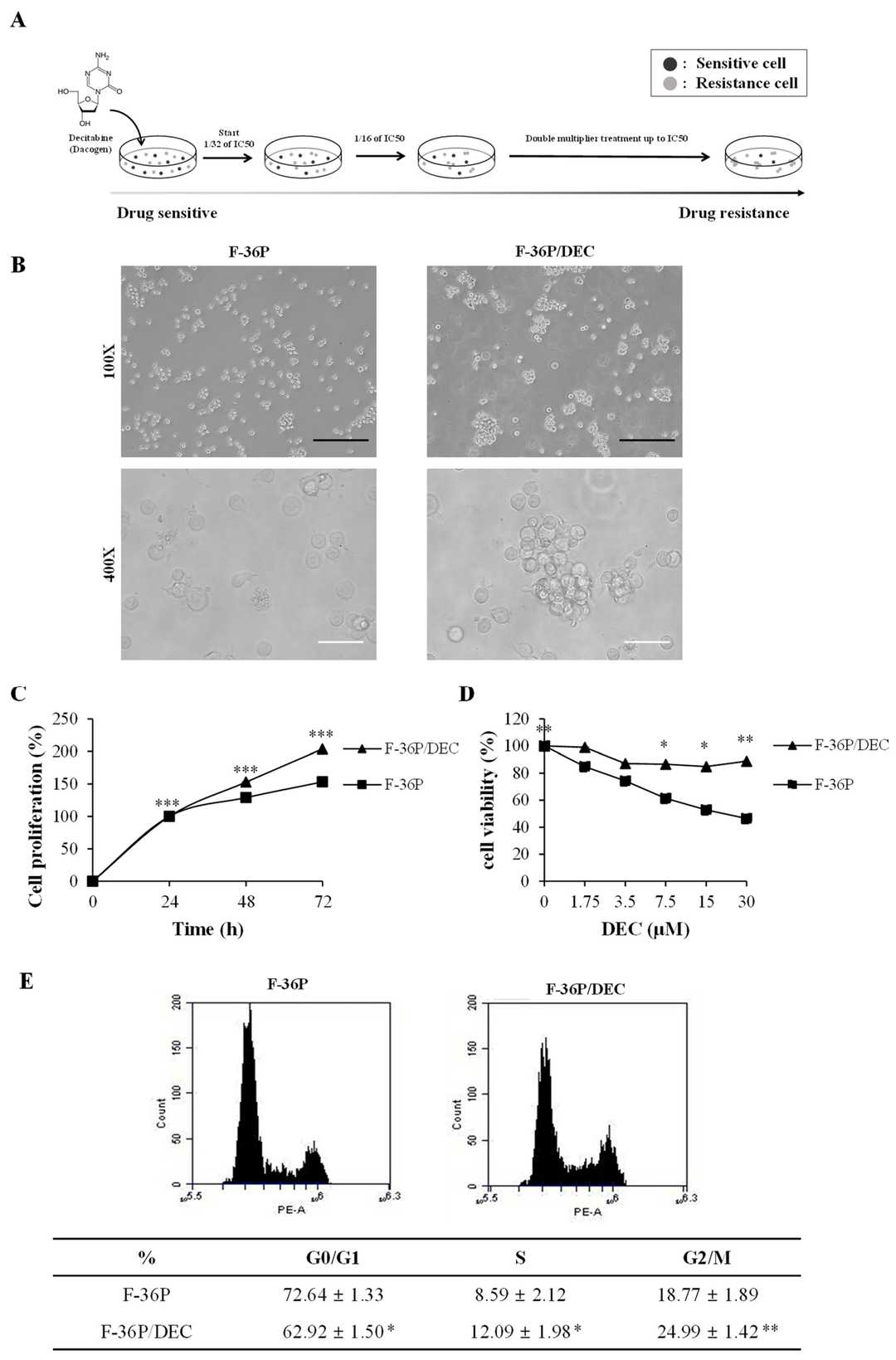 Fig. 3. Validation of DEC resistance in the F-36P/DEC cell line (Kim S, Shin D-Y, et al., 2021).
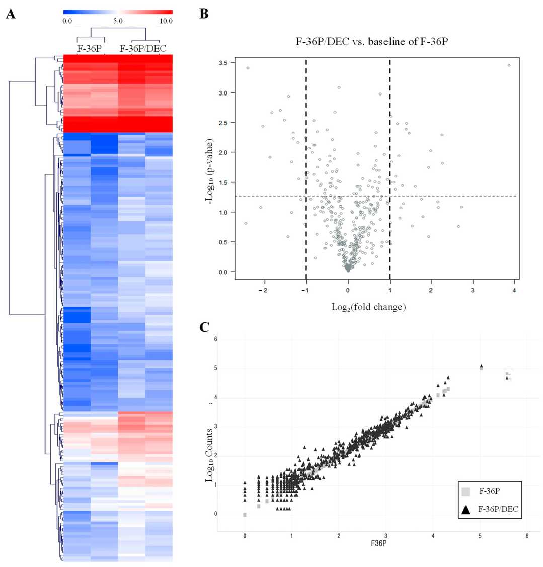 Fig. 4. Analysis of DEGs between F-36P/DEC and F-36P cells (Kim S, Shin D-Y, et al., 2021).
Ask a Question
Write your own review
- You May Also Need

Description: Established in 2007 from the bone marrow mononuclear cells of an 82-year-old Japanese man with diffuse large B-cell lymphoma in the leukemic phase

Description: Established from the bone marrow of a 28-year-old man who developed the terminal leukemic phase of lymphosarcoma in 1976

Description: This cell line was derived from the bone marrow aspirate of a 59 year old male with erythroleukemia that became acute myelogenous leukaemia.The cells form colonies in soft-agar in the presence of ...

Description: Established from the pleural effusion of a 24-year-old woman with recurrent anaplastic large cell lymphoma (ALCL); cells were described to clonally derive from T-lineage lymphoid cells and to be ...

Description: Established from a 37-year-old man at second (refractory/terminal) relapse of Hodgkin lymphoma (nodular sclerosing -> lymphocyte depleted/stage IIISA -> stage IV) after both combined chemo- and ...

Description: Established from the peripheral blood of a 10-year-old Caucasian boy with acute lymphoblastic leukemia (pre B-ALL) at diagnosis in 1993
- Adipose Tissue-Derived Stem Cells
- Human Neurons
- Mouse Probe
- Whole Chromosome Painting Probes
- Hepatic Cells
- Renal Cells
- In Vitro ADME Kits
- Tissue Microarray
- Tissue Blocks
- Tissue Sections
- FFPE Cell Pellet
- Probe
- Centromere Probes
- Telomere Probes
- Satellite Enumeration Probes
- Subtelomere Specific Probes
- Bacterial Probes
- ISH/FISH Probes
- Exosome Isolation Kit
- Human Adult Stem Cells
- Mouse Stem Cells
- iPSCs
- Mouse Embryonic Stem Cells
- iPSC Differentiation Kits
- Mesenchymal Stem Cells
- Immortalized Human Cells
- Immortalized Murine Cells
- Cell Immortalization Kit
- Adipose Cells
- Cardiac Cells
- Dermal Cells
- Epidermal Cells
- Peripheral Blood Mononuclear Cells
- Umbilical Cord Cells
- Monkey Primary Cells
- Mouse Primary Cells
- Breast Tumor Cells
- Colorectal Tumor Cells
- Esophageal Tumor Cells
- Lung Tumor Cells
- Leukemia/Lymphoma/Myeloma Cells
- Ovarian Tumor Cells
- Pancreatic Tumor Cells
- Mouse Tumor Cells