
C-33A
Cat.No.: CSC-C9090W
Species: Homo sapiens (Human)
Source: Uterus; Cervix
Morphology: Epithelial
- Specification
- Background
- Scientific Data
- Q & A
- Customer Review
C-33A is a well-characterized human cell line derived from a cervical carcinoma. Originally established from a poorly differentiated squamous cell carcinoma of the cervix, this adherent epithelial line exhibits typical polygonal morphology and robust growth in standard culture conditions.
A defining and scientifically valuable feature of C-33A is its confirmed lack of human papillomavirus (HPV) sequences. Unlike the majority of cervical cancer cell lines (e.g., HeLa, SiHa, CaSki), which harbor integrated HPV genomes, C-33A is HPV-negative. This property renders it an indispensable negative control for investigating HPV-dependent versus HPV-independent pathways in cervical carcinogenesis, including p53 and retinoblastoma protein (pRb) regulation. Notably, C-33A carries a homozygous missense mutation in the TP53 gene (codon 273, Arg→His), leading to expression of a mutant p53 protein. This mutation further distinguishes it from HPV-positive lines that inactivate wild-type p53 through E6-mediated degradation.
The major advantage of C-33A lies in its utility for functional studies of oncogenic mechanisms unrelated to viral oncoproteins. It enables researchers to assess the intrinsic effects of genetic manipulations, drug responses, and signaling pathways without interference from HPV E6/E7. Additionally, C-33A is widely employed in chemosensitivity assays, radiotherapy research, and comparative genomic analyses. Its ease of transfection and reproducible behavior make it a robust model for cervical cancer biology, particularly for exploring non-HPV-driven tumor progression and therapeutic targets.
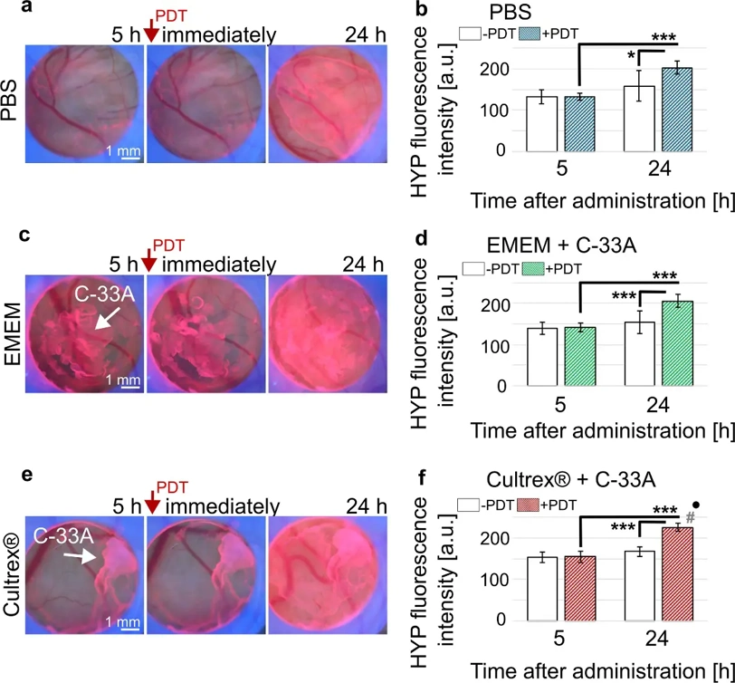
Establishment of C-33A Cervical Carcinoma Spheroids on Japanese Quail CAM
Cervical cancer is the most common type of cancer in women. The present study aims to establish a reliable model of human cervical cancer microtumours (C-33A) growing on the chorioallantoic membrane (CAM) of quail embryos to test the efficacy of anticancer drugs. Hypericin (HYP), a natural perylene quinone, was used in this study as a model photosensitizer for photodiagnosis and photodynamic therapy (PDT). Fluorescence pharmacokinetics were recorded to verify the robustness of the model. The changes in vessel growth parameters were determined after weak PDT (405 nm, 2 min and 285 mW/cm2) with the aim to reduce vascularization and nutrition of the C-33A spheroids. Vasoconstriction and occlusion of small vessels and capillaries were identified in the CAM. Subsequently, histological examination revealed changes in the chorioallantoic ectoderm of the CAM. An invasion of C-33A cells into the CAM was confirmed. Furthermore, increased expression of growth factor genes was detected by qPCR, indicating angiogenesis of the CAM tissue. In summary, C-33A cells grown on CAM were shown as a valuable 3D spheroid model for photodiagnosis and PDT assays of cervical cancer.
Ask a Question
Write your own review
- You May Also Need


- Adipose Tissue-Derived Stem Cells
- Human Neurons
- Mouse Probe
- Whole Chromosome Painting Probes
- Hepatic Cells
- Renal Cells
- In Vitro ADME Kits
- Tissue Microarray
- Tissue Blocks
- Tissue Sections
- FFPE Cell Pellet
- Probe
- Centromere Probes
- Telomere Probes
- Satellite Enumeration Probes
- Subtelomere Specific Probes
- Bacterial Probes
- ISH/FISH Probes
- Exosome Isolation Kit
- Human Adult Stem Cells
- Mouse Stem Cells
- iPSCs
- Mouse Embryonic Stem Cells
- iPSC Differentiation Kits
- Mesenchymal Stem Cells
- Immortalized Human Cells
- Immortalized Murine Cells
- Cell Immortalization Kit
- Adipose Cells
- Cardiac Cells
- Dermal Cells
- Epidermal Cells
- Peripheral Blood Mononuclear Cells
- Umbilical Cord Cells
- Monkey Primary Cells
- Mouse Primary Cells
- Breast Tumor Cells
- Colorectal Tumor Cells
- Esophageal Tumor Cells
- Lung Tumor Cells
- Leukemia/Lymphoma/Myeloma Cells
- Ovarian Tumor Cells
- Pancreatic Tumor Cells
- Mouse Tumor Cells