
NB9
Cat.No.: CSC-C6501J
Species: Homo sapiens (Human)
Morphology: other
Culture Properties: Adherent/Suspension cells
- Specification
- Background
- Scientific Data
- Q & A
- Customer Review
Store in liquid nitrogen.
NB9 (also NB-9 or NB9) is a cancer cell line derived from a human neuroblastoma, a solid tumor that affects the sympathetic nervous system. Neuroblastoma most often originates from the adrenal medulla or sympathetic chain and is most common in children. NB9 was developed from the primary tumor tissue of a 5-year-old female neuroblastoma patient. NB9 is tumorigenic and immortalized. NB9, like other neuroblastoma-derived cell lines, has the potential to display variable morphology and expression of tumor-associated genes. NB9 is frequently used to study neuroblastoma pathogenesis and development. NB9 has been used in several studies published on the genetics behind neuroblastic diseases including RAS-related and other oncogenic mutations. NB9 cells may be cultured under typical mammalian tissue culture conditions and maintained in serum-supplemented media (e.g. RPMI-1640 with fetal bovine serum). NB9 cells have properties that allow them to be cultured in suspension or as an adherent monolayer, depending on the application. This cell line is a model for drug screening and pharmacologic testing, genetic perturbation, signaling studies, and other applications related to the study of pediatric neuroblastoma and putative therapeutic interventions.
Effects of Dedifferentiated Fat Cells on Neurogenic Differentiation and Cell Proliferation in Neuroblastoma Cells
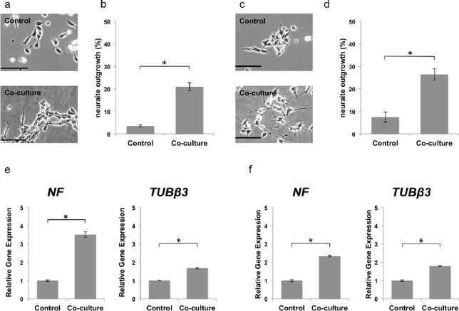
Mesenchymal stem cells (MSCs) can induce neuroblastoma (NB) cell differentiation. Dedifferentiated fat cells (DFATs) share properties with MSCs. Here, Hidaka et al. cultured NB cells with DFATs and DFAT-conditioned medium (CM) with or without a PI3K inhibitor, aiming to investigate whether DFATs can induce NB cell differentiation and suppress proliferation.
When observing NB9 cell morphology with a phase-contrast microscope, the co-culture group had more cells with neurites than the control group (Fig. 1a). Quantitative analysis showed a significantly higher percentage of cells with neurite outgrowth in the co-culture group (Fig. 1b). SK-N-SH cells also exhibited similar results (Fig. 1c and d). After confirming NB9 cell differentiation through neurite outgrowth assays, real-time RT-PCR was used to measure mRNA expression of neuronal-specific markers (NF and TUBβ3). Both NB9 and SK-N-SH cells in the co-culture group had significantly higher mRNA levels of NF and TUBβ3 compared to their respective controls (Fig. 1e and f).
Ask a Question
Write your own review
- You May Also Need

Description: Subline of GOTO. Protein-free medium adapted. Cell growth is slow.


Description: Established from the adrenal tumor tissue resected after treatment from a 20-month-old boy of European origin with neuroblastoma (stage III) in 1991
Description: established from the bone marrow metastasis of a 1-year-old female patient with adrenal gland neuroblastoma negative

Description: The line is resistant to infection and focus formation by ecotropic MuLV.
Description: Established from the primary adrenal tumor resected prior to treatment from a 3-year-old boy with rapidly progressing Stage III neuroblastoma in 1989; described to lack MYCN (NMYC, N-myc) ...
- Adipose Tissue-Derived Stem Cells
- Human Neurons
- Mouse Probe
- Whole Chromosome Painting Probes
- Hepatic Cells
- Renal Cells
- In Vitro ADME Kits
- Tissue Microarray
- Tissue Blocks
- Tissue Sections
- FFPE Cell Pellet
- Probe
- Centromere Probes
- Telomere Probes
- Satellite Enumeration Probes
- Subtelomere Specific Probes
- Bacterial Probes
- ISH/FISH Probes
- Exosome Isolation Kit
- Human Adult Stem Cells
- Mouse Stem Cells
- iPSCs
- Mouse Embryonic Stem Cells
- iPSC Differentiation Kits
- Mesenchymal Stem Cells
- Immortalized Human Cells
- Immortalized Murine Cells
- Cell Immortalization Kit
- Adipose Cells
- Cardiac Cells
- Dermal Cells
- Epidermal Cells
- Peripheral Blood Mononuclear Cells
- Umbilical Cord Cells
- Monkey Primary Cells
- Mouse Primary Cells
- Breast Tumor Cells
- Colorectal Tumor Cells
- Esophageal Tumor Cells
- Lung Tumor Cells
- Leukemia/Lymphoma/Myeloma Cells
- Ovarian Tumor Cells
- Pancreatic Tumor Cells
- Mouse Tumor Cells