
U266B1
Cat.No.: CSC-C9138W
Species: Homo sapiens (Human)
Source: Blood; Peripheral Blood
Morphology: round to polygonal
- Specification
- Background
- Scientific Data
- Q & A
- Customer Review
U266B1, also referred to as U-266, is an established human multiple myeloma cell line. It was originally derived from the peripheral blood of a 53-year-old male patient with IgE-producing myeloma. As their name suggests, U266B1 cells are lymphoblast-like in morphology. They proliferate almost exclusively in suspension with minimal attachment to substrata. These cells secrete the cytokine interleukin-6 (IL-6) and immunoglobulin E (IgE) with λ light chains. These cells express classic plasma cell-associated antigens, including CD38, CD44, CD56, syndecan-1 (CD138) and BCMA, while they do not express classical B-cell antigens, such as CD19 and CD20.
Functionally, U266B1 cells exhibit autocrine signaling through IL-6, as these cells grow without exogenous IL-6. The doubling time of these cells has been reported to be approximately 55-144 hours depending on culture conditions. These cells are tumorigenic in vivo, as they engraft and proliferate in immunocompromised mice in either the bone marrow compartment (when given systemically) or as subcutaneous xenografts. The U266B1 cell line is used in multiple myeloma research to model malignant plasma cell biology, IL-6-mediated growth and survival, mechanisms of drug resistance and tumor-microenvironment interactions. U266B1 has also been used as a fusion partner to produce human monoclonal antibodies using hybridoma technology.
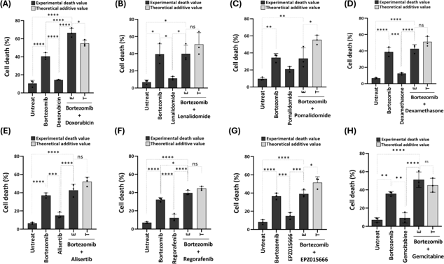
Doxorubicin Synergized Bortezomib-Induced Cell Death in U266B1 Cells
Bortezomib is a standard treatment for multiple myeloma (MM) but faces resistance due to aggresome formation, which sequesters misfolded proteins. Here, Yu et al. screened anticancer compounds in U266B1 MM cells to enhance bortezomib efficacy.
They screened various anticancer agents for their potential to enhance bortezomib-induced cell death in the MM cell line U266B1. This included gemcitabine, regorafenib, lenalidomide, pomalidomide, dexamethasone, doxorubicin, EPZ01566, and alisertib. Bortezomib at 6.25 nM caused ~30-40% cell death. Test compounds, each inducing 10-20% cell death, were used alone or with bortezomib. Cell viability was assessed 24 h later using the trypan blue exclusion assay. Results showed that only doxorubicin produced synergistic cell death with bortezomib, evidenced by a significantly higher observed cell death rate (E) compared to the sum of the cell death rates from bortezomib and doxorubicin (T) (Fig. 1). Other compounds either produced an additive effect (E ≈ T) or failed to achieve an additive effect (E < T).
Ask a Question
Write your own review
- You May Also Need
Description: Established in 2007 from the large retrosternal mass resected before treatment from a 57-year-old Caucasian man with rapidly fatal anaplastic thyroid cancer

Description: established from the uvea tissue of a male patient with uveal melanoma

Description: NTERA-2 was cloned from cell line TERA-2 which was derived from a metastatic teratocarcinoma of a 22-year-old Caucasian male; cell line also known as NT-2
Description: Established from the bone marrow of a 27-year-old woman with B-cell non-Hodgkin lymphoma (B-NHL) (diffuse large cell lymphoma, DLCL, stage 4B, at relapse) in 1987
- Adipose Tissue-Derived Stem Cells
- Human Neurons
- Mouse Probe
- Whole Chromosome Painting Probes
- Hepatic Cells
- Renal Cells
- In Vitro ADME Kits
- Tissue Microarray
- Tissue Blocks
- Tissue Sections
- FFPE Cell Pellet
- Probe
- Centromere Probes
- Telomere Probes
- Satellite Enumeration Probes
- Subtelomere Specific Probes
- Bacterial Probes
- ISH/FISH Probes
- Exosome Isolation Kit
- Human Adult Stem Cells
- Mouse Stem Cells
- iPSCs
- Mouse Embryonic Stem Cells
- iPSC Differentiation Kits
- Mesenchymal Stem Cells
- Immortalized Human Cells
- Immortalized Murine Cells
- Cell Immortalization Kit
- Adipose Cells
- Cardiac Cells
- Dermal Cells
- Epidermal Cells
- Peripheral Blood Mononuclear Cells
- Umbilical Cord Cells
- Monkey Primary Cells
- Mouse Primary Cells
- Breast Tumor Cells
- Colorectal Tumor Cells
- Esophageal Tumor Cells
- Lung Tumor Cells
- Leukemia/Lymphoma/Myeloma Cells
- Ovarian Tumor Cells
- Pancreatic Tumor Cells
- Mouse Tumor Cells