
OS-RC-2
Cat.No.: CSC-C9232W
Species: Homo sapiens (Human)
Source: kidney
Morphology: epithelial-like
- Specification
- Background
- Scientific Data
- Q & A
- Customer Review
The OS-RC-2 cell line is a well-established human renal cell carcinoma (RCC) model derived from a primary clear cell RCC (ccRCC) tumor of a Chinese patient. OS-RC-2 cells exhibit an epithelial morphology and carry the hallmark genetic alterations of ccRCC. They possess biallelic von Hippel-Lindau (VHL) gene inactivation, leading to constitutive stabilization of hypoxia-inducible factors (HIF-1α and HIF-2α) even under normoxic conditions.
The primary utility of the OS-RC-2 cell line stems from its status as a VHL-deficient, HIF-active ccRCC model with direct human tumor origin. It serves as a functional tool to study the consequences of VHL loss and the downstream roles of HIF-α isoforms in driving ccRCC progression. This makes it directly relevant for testing therapies that target HIF-mediated transcription or its downstream effectors (e.g., VEGF inhibitors).
Carbonic Anhydrase IX-Targeted Molecular Probe for PET Imaging of Clear Cell Renal Cell Carcinoma
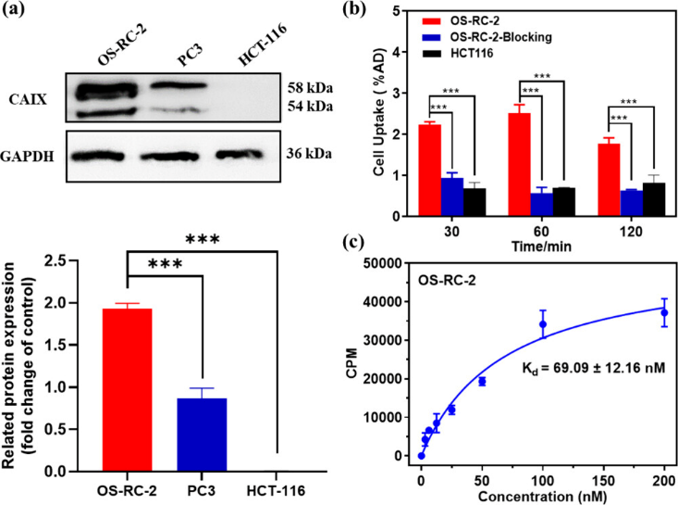
Carbonic anhydrase IX (CAIX) has been considered as a promising biomarker for diagnosing and treating clear cell renal cell carcinoma (ccRCC). Therefore, sensitive and accurate detection of the CAIX expression level is crucial for guiding therapeutic decisions and prognostic assessment of ccRCC. In this study, a small molecular imaging probe, 18F-DAZ, was designed and synthesized for specifically and sensitively visualizing CAIX expression.
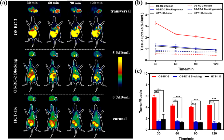
18F-DAZ exhibited high radiochemical purity (>98%), high stability, and high affinity for CAIX-positive cancer cells OS-RC-2 with the Kd value of 69.09 ± 12.16 nM. The probe uptake in OS-RC-2 cells was higher than that in the OS-RC-2 blocking group and CAIX-negative cells HCT-116 at the same time. Positron emission tomography (PET) imaging results showed that the highest accumulation of the probe in OS-RC-2 bearing mice was also markedly higher than that in the OS-RC-2 blocking group and HCT-116. Biodistribution and autoradiography analysis further verified that the probe 18F-DAZ can specifically target CAIX. These results highlight the potential of 18F-DAZ as a candidate PET imaging radiotracer for detecting CAIX-overexpressing tumors.
Ask a Question
Write your own review
- You May Also Need


Description: Rhabdoid tumor of kidney (formerly classified as Wilms' tumor)


Description: The cells express the transforming gene of adenovirus 5. Adenovirus 5 DNA from both the right and left ends of the viral genome are present. The line is excellent for titrating human adenoviruses. ...
- Adipose Tissue-Derived Stem Cells
- Human Neurons
- Mouse Probe
- Whole Chromosome Painting Probes
- Hepatic Cells
- Renal Cells
- In Vitro ADME Kits
- Tissue Microarray
- Tissue Blocks
- Tissue Sections
- FFPE Cell Pellet
- Probe
- Centromere Probes
- Telomere Probes
- Satellite Enumeration Probes
- Subtelomere Specific Probes
- Bacterial Probes
- ISH/FISH Probes
- Exosome Isolation Kit
- Human Adult Stem Cells
- Mouse Stem Cells
- iPSCs
- Mouse Embryonic Stem Cells
- iPSC Differentiation Kits
- Mesenchymal Stem Cells
- Immortalized Human Cells
- Immortalized Murine Cells
- Cell Immortalization Kit
- Adipose Cells
- Cardiac Cells
- Dermal Cells
- Epidermal Cells
- Peripheral Blood Mononuclear Cells
- Umbilical Cord Cells
- Monkey Primary Cells
- Mouse Primary Cells
- Breast Tumor Cells
- Colorectal Tumor Cells
- Esophageal Tumor Cells
- Lung Tumor Cells
- Leukemia/Lymphoma/Myeloma Cells
- Ovarian Tumor Cells
- Pancreatic Tumor Cells
- Mouse Tumor Cells