OCI-LY-1
Cat.No.: CSC-C9227W
Species: Homo sapiens (Human)
Source: Bone Marrow
Morphology: small round to polymorph cells
- Specification
- Background
- Scientific Data
- Q & A
- Customer Review
The OCI-LY-1 cell line is a pivotal resource in the study of B-cell non-Hodgkin lymphoma (B-NHL), providing invaluable insights into the disease's pathology, genetic basis, and potential therapeutic targets. Established in 1983 from the bone marrow of a 44-year-old man undergoing a relapse of stage 4B diffuse large B-cell lymphoma (DLBCL), OCI-LY-1 cells encapsulate the aggressive nature and complex biology of this lymphoma subtype.
DLBCL, the subtype represented by the OCI-LY-1 cell line, is recognized for its heterogeneity, both clinically and molecularly. This cell line has been assigned to the germinal center B-cell (GCB) lymphoma subtype, which is one of the primary subtypes of DLBCL identified through gene expression profiling. The GCB subtype originates from B-cells within the germinal center and is characterized by specific genetic and molecular features that differentiate it from other DLBCL subtypes. This distinction is crucial, as it influences the response to treatment and overall prognosis.
The derivation and maintenance of the OCI-LY-1 cell line have made it a cornerstone for numerous studies aimed at uncovering the pathophysiological mechanisms underlying B-NHL, especially the GCB subtype of DLBCL. Researchers utilize OCI-LY-1 cells to investigate genetic mutations, signaling pathways, and the microenvironment interactions that drive lymphomagenesis. Furthermore, through the study of OCI-LY-1 cells, significant progress has been made in understanding the molecular characteristics of GCB-like DLBCL, leading to the identification of potential biomarkers for diagnosis and treatment stratification.
Molecular Cytogenetic Characterization of NHL Cell Lines
Spectral karyotyping (SKY) and comparative genomic hybridization (CGH) have greatly enhanced the resolution of cytogenetic analysis, enabling the identification of novel regions of rearrangement and amplification in tumor cells. 10 malignant non-Hodgkin lymphoma (NHL) cell lines, including OCI-Ly1, OCI-Ly2, OCI-Ly3, OCI-LY4, OCI-Ly7, OCI-Ly8, OCI-Ly12, OCI-Ly13.2, OCI-Ly17, and OCI-Ly18, were analyzed by G-banding, SKY, and CGH. Fig. 1 illustrates the display and classified SKY images of a representative metaphase cell from OCI-Ly17 after hybridization with SKY painting probes.
In contrast to the 52 breakpoints identified by G-banding, SKY identified 87 breakpoints, which clustered at 1q21, 7p15, 8p11, 13q21, 13q32, 14q32, 17q11, and 18q21. G-banding identified 10 translocations, including the previously described recurring translocations, t(8;14)(q24;q32) and t(14;18)(q32;q21). In contrast, SKY identified 60 translocations, including five that were recurring, t(8;14)(q24;q32), t(14;18)(q32;q21), t(4;7)(p12;q22), t(11;18)(q22;q21), and t(3;18)(q21;p11) (Fig. 2). CGH identified seven sites of high-level DNA amplification, 1q31-32, 2p12-16, 8q24, 11q23-25, 13q21-22, 13q32-34, and 18q21-23; of these, 1q31-32, 11q23-25, 13q21-22, and 13q32-34 have previously not been described as amplified in NHL.
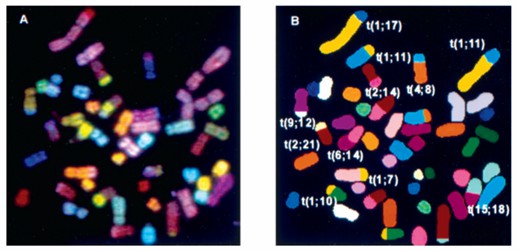 Fig. 1 A metaphase cell from the cell line OCI-Ly17 showing display (A) and classification (B) colors. (Mehra S, et al., 2002)
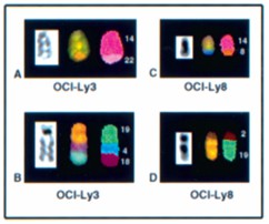 Fig. 2 Representative examples of rearrangements misidentified by G-banding that SKY identified. (Mehra S, et al., 2002)
Effects of Different Concentrations of Doxorubicin on OCI-LY1 Cells
Adriamycin (doxorubicin) is an important traditional drug that exhibits cytotoxicity in DLBCL. At the concentration of 8 μmol/l of doxorubicin, the inhibition rate of OCI-LY1 cell proliferation was found to be 69.313 ± 5.359%, which was significantly higher than the inhibition rates 59.427 ± 4.145%, 39.563 ± 7.350%, 25.340 ± 4.835%, and 17.108 ± 3.957% at 4, 2, 1, and 0.5 μmol/l concentrations, respectively (P < 0.05). An increase in the concentration of doxorubicin resulted in a gradual increase in the suppression of the proliferation of OCI-LY1 cells, with significant differences between the groups (P < 0.05). The IC50 of doxorubicin used to intervene with OCI-LY1 cells was found to be 3.028 μmol/l for 24 h, which laid a foundation for future experiments. Overall, doxorubicin hindered the proliferation of DLBCL cells, with an increase in the concentration of doxorubicin enhancing the inhibition rate of DLBCL cells (Fig. 3 and 4).
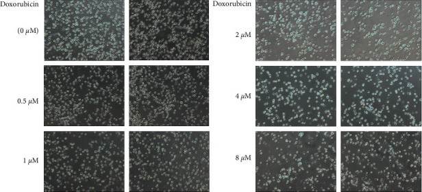 Fig. 3 Images of doxorubicin interfering with the proliferation of OCI-LY1 cells. (Yan S, et al., 2022)
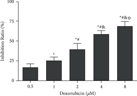 Fig. 4 Effect of doxorubicin on the proliferation of OCI-LY1 cells. (Yan S, et al., 2022)
Ask a Question
Write your own review
- You May Also Need
Description: Established in 2007 from the large retrosternal mass resected before treatment from a 57-year-old Caucasian man with rapidly fatal anaplastic thyroid cancer

Description: established from the uvea tissue of a male patient with uveal melanoma

Description: NTERA-2 was cloned from cell line TERA-2 which was derived from a metastatic teratocarcinoma of a 22-year-old Caucasian male; cell line also known as NT-2

Description: Established from the bone marrow of a 27-year-old woman with B-cell non-Hodgkin lymphoma (B-NHL) (diffuse large cell lymphoma, DLCL, stage 4B, at relapse) in 1987
- Adipose Tissue-Derived Stem Cells
- Human Neurons
- Mouse Probe
- Whole Chromosome Painting Probes
- Hepatic Cells
- Renal Cells
- In Vitro ADME Kits
- Tissue Microarray
- Tissue Blocks
- Tissue Sections
- FFPE Cell Pellet
- Probe
- Centromere Probes
- Telomere Probes
- Satellite Enumeration Probes
- Subtelomere Specific Probes
- Bacterial Probes
- ISH/FISH Probes
- Exosome Isolation Kit
- Human Adult Stem Cells
- Mouse Stem Cells
- iPSCs
- Mouse Embryonic Stem Cells
- iPSC Differentiation Kits
- Mesenchymal Stem Cells
- Immortalized Human Cells
- Immortalized Murine Cells
- Cell Immortalization Kit
- Adipose Cells
- Cardiac Cells
- Dermal Cells
- Epidermal Cells
- Peripheral Blood Mononuclear Cells
- Umbilical Cord Cells
- Monkey Primary Cells
- Mouse Primary Cells
- Breast Tumor Cells
- Colorectal Tumor Cells
- Esophageal Tumor Cells
- Lung Tumor Cells
- Leukemia/Lymphoma/Myeloma Cells
- Ovarian Tumor Cells
- Pancreatic Tumor Cells
- Mouse Tumor Cells