- Specification
- Background
- Scientific Data
- Q & A
- Customer Review
The NCCIT cell line originates from the teratoma component of a mixed germ cell tumor located in the anterior mediastinum of a 24-year-old man and was established by Dr. Shinichi Teshima at the National Cancer Center Research Institute in Tokyo, Japan during 1985. Anterior mediastinal mixed germ cell tumors represent rare neoplasms that possess multiple germ cell types such as spermatogonia along with yolk sac and trophoblastic cells. The tumor displays both embryonal carcinoma and seminoma features and exhibits substantial heterogeneity.
NCCIT cells possess significant differentiation potential, being able to differentiate into embryonic stem cell-like cells under retinoic acid induction, and they express embryonic and pluripotency markers such as OCT4, SOX2, and NANOG. Genomic reprogramming occurs during the differentiation process which leads to various epigenetic modifications including dynamic DNA methylation and histone modification changes. Scientists utilize NCCIT cells for research in germ cell tumor pathogenesis and cancer stem cell biology because these cells possess pluripotency and self-renewal capabilities. For example, studies demonstrate the essential function of the Wnt/β-catenin signaling pathway in initiating tumor development within NCCIT cells. Scientists use NCCIT cells as a model to evaluate anticancer drug efficacy. Studies reveal that tannic acid initiates programmed cell death in NCCIT cells by altering ROS concentrations which results in suppressed cell growth. Scientists employ NCCIT cells for research on cisplatin resistance mechanisms and genetic mutation effects.
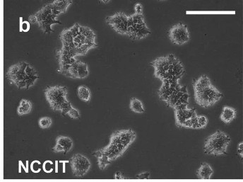 Fig. 1. Morphology of NCCIT cell line, Scalebar: 500 μm (Jostes S, Nettersheim D, et al., 2021).
TA Inhibits Cell Proliferation of NCCIT Cells as Well as Cancer Stem Cell Markers
The shared gene expression patterns between human embryonic carcinoma cells and embryonic stem cells lead to abnormal self-renewal abilities and tumor formation in breast cancer. The Wnt/β-catenin pathway plays a vital role in maintaining cancer stem cell viability and phytochemicals like tannic acid can inhibit this pathway. Sp et al. investigated how tannic acid activates TRAIL-mediated cell death in human embryonic carcinoma cells through mitochondrial reactive oxygen species control which may lead to enhanced cancer treatment methods.
A crystal violet assay was used to determine how TA affects NCCIT cell proliferation by comparing TA-treated cells to untreated control cells (Fig. 1A). Results demonstrated that TA inhibited cell proliferation according to its concentration after both 24 and 48 hours. Research used the 50 μM concentration identified as the IC50 after 48 hours for subsequent experiments. The effect of TA on stem cell markers in NCCIT cells was examined by measuring mRNA levels of SOX2, OCT4, and NANOG which displayed a considerable decrease due to TA treatment (Fig. 2A and B). Real-time PCR analysis verified the inhibition as shown in Figure S1B. Analysis of protein levels demonstrated that TA led to a significant decrease in the markers SOX2, OCT4, and NANOG as indicated by Figure 2C and D.
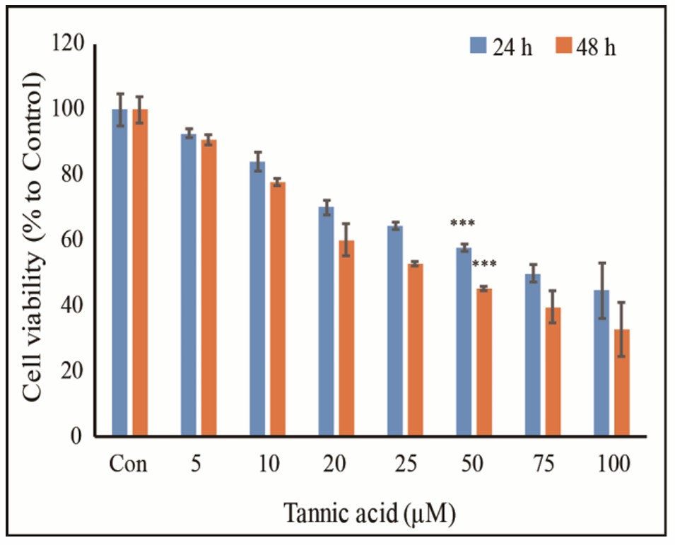 Fig. 1. TA inhibits NCCIT cell proliferation (Sp N, Kang DY, et al., 2020).
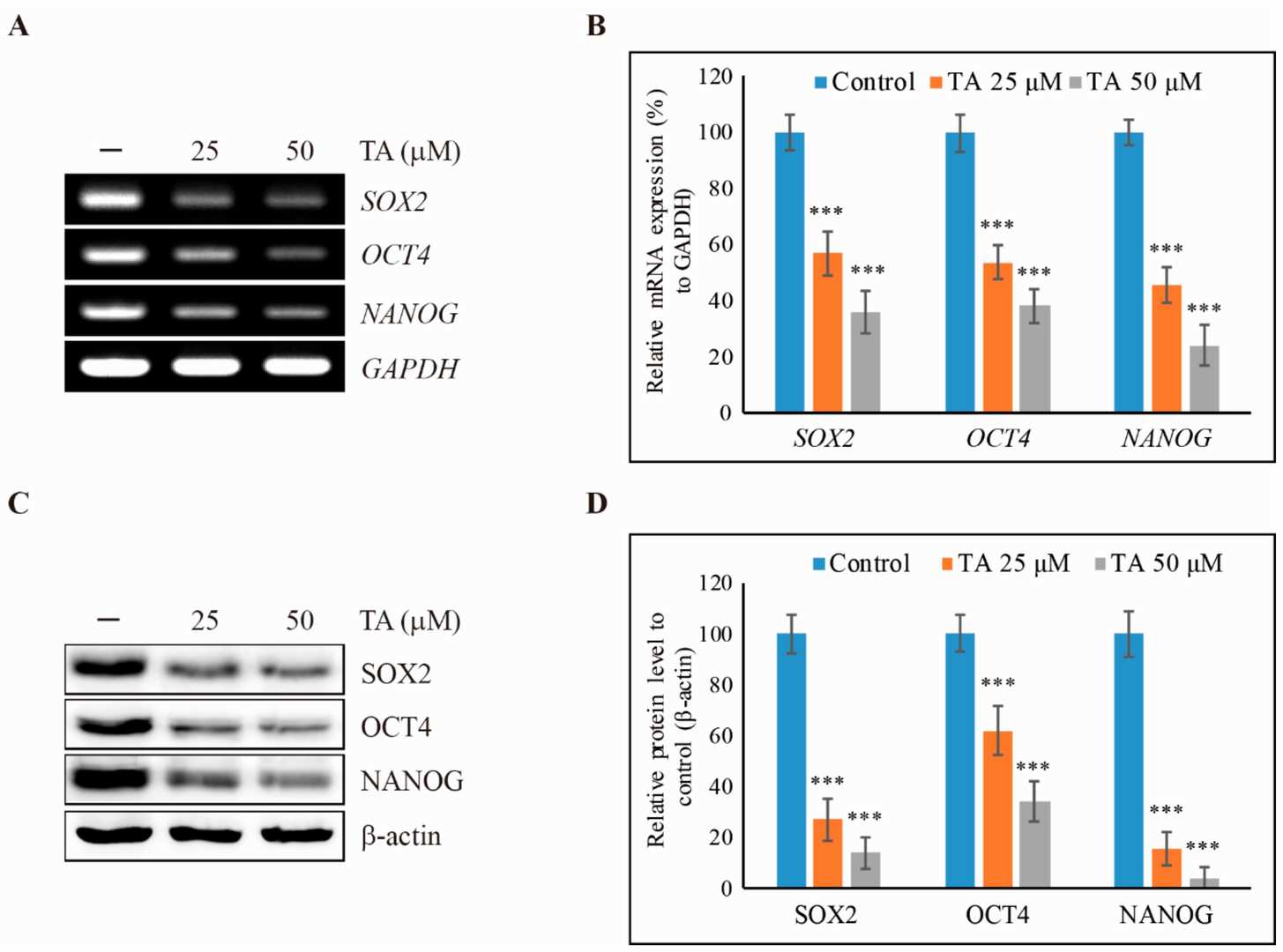 Fig. 2. Tannic acid (TA) inhibits cancer stem cell markers in NCCIT cells (Sp N, Kang DY, et al., 2020).
Mediastinal GCT Cell Line NCCIT Harbors Low Levels of MDM2 and Mutant TP53 whereas Testicular GCT Cell Line 2102Ep Harbors Wild-Type TP53 and High Levels of MDM2
GCTs are highly treatable, yet mediastinal variants exhibit poor responsiveness possibly due to TP53 mutations. To investigate differences between mediastinal and testicular GCTs, Timmerman et al. utilized NCCIT and 2102Ep cell lines. The 2102Ep line is derived from testis, while NCCIT originates from the mediastinum, both having a similar differentiation state. The 2102Ep line has a wild-type TP53, whereas NCCIT features a hemizygous deletion causing a frameshift and premature stop codon (Fig. 3A), consistent with TP53 mutations in mediastinal GCTs.
They used isogenic clones of NCCIT and 2102Ep with acquired cisplatin resistance from prolonged sublethal exposure (see Materials and Methods). RNA sequencing revealed lower MDM2 expression in both NCCIT lines compared to 2102Ep, with resistant 2102Ep showing the highest MDM2 levels (Fig. 3B), confirmed by Western blot (Fig. 3C). MDM4 levels were similar across all lines (Fig. 4). Principal component analysis showed close similarities between parental and resistant subclones. Despite low MDM2 levels, NCCIT cells treated with sublethal cisplatin showed decreased MDM2 and MDM4 levels, indicating a functional DNA damage response and intact P53 regulation, contrary to the expected null TP53 status (Fig. 3D and E).
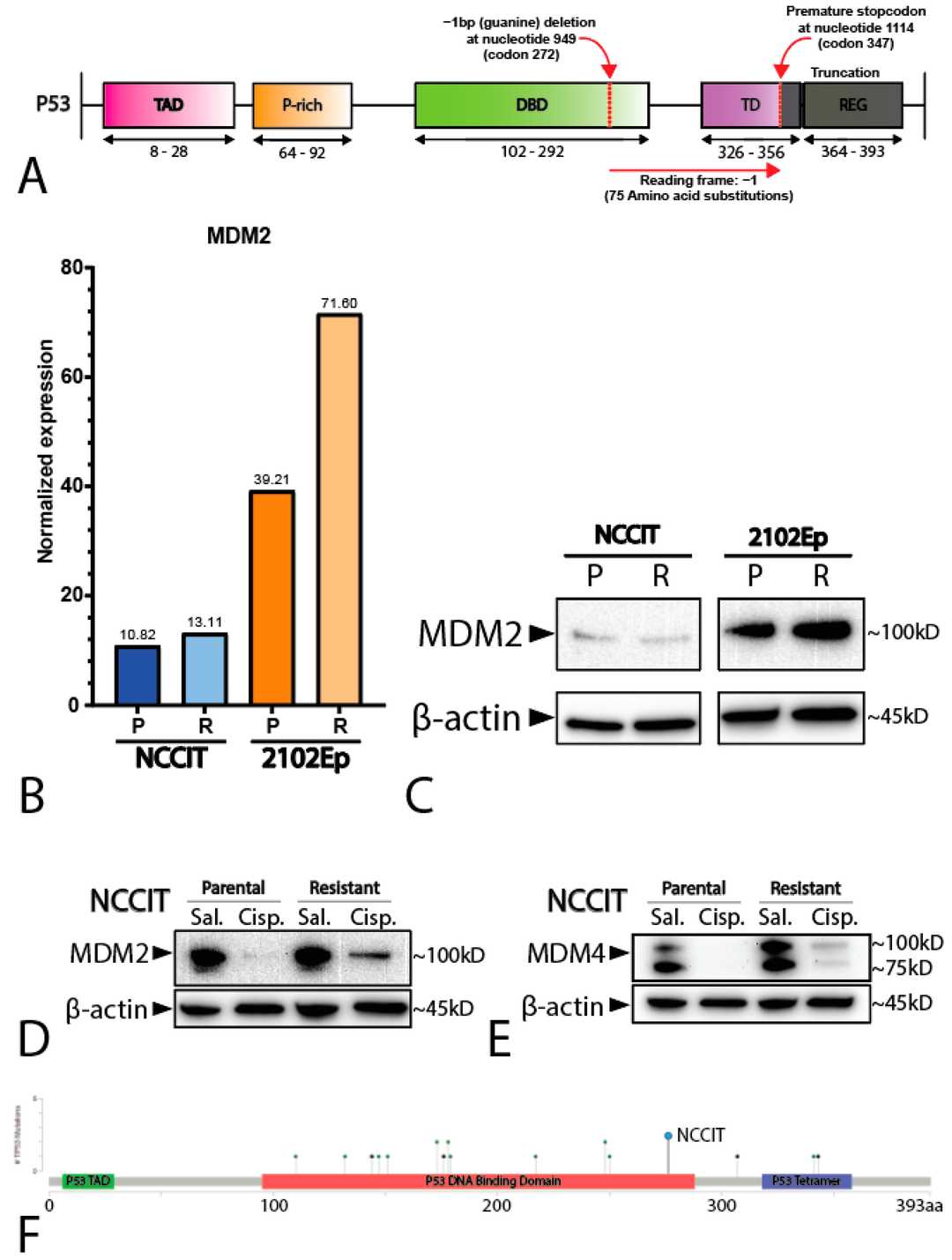 Fig. 3. Characterization of the cell lines NCCIT and 2102Ep (Timmerman DM, Eleveld TF, et al., 2021).
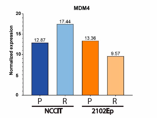 Fig. 4. Normalized expression of MDM4 (RNAseq) in NCCIT and 2102Ep (both parental and resistant) (Timmerman DM, Eleveld TF, et al., 2021).
Ask a Question
Write your own review
- You May Also Need
Description: Established in 2007 from the large retrosternal mass resected before treatment from a 57-year-old Caucasian man with rapidly fatal anaplastic thyroid cancer

Description: established from the uvea tissue of a male patient with uveal melanoma

Description: NTERA-2 was cloned from cell line TERA-2 which was derived from a metastatic teratocarcinoma of a 22-year-old Caucasian male; cell line also known as NT-2
Description: Established from the bone marrow of a 27-year-old woman with B-cell non-Hodgkin lymphoma (B-NHL) (diffuse large cell lymphoma, DLCL, stage 4B, at relapse) in 1987
- Adipose Tissue-Derived Stem Cells
- Human Neurons
- Mouse Probe
- Whole Chromosome Painting Probes
- Hepatic Cells
- Renal Cells
- In Vitro ADME Kits
- Tissue Microarray
- Tissue Blocks
- Tissue Sections
- FFPE Cell Pellet
- Probe
- Centromere Probes
- Telomere Probes
- Satellite Enumeration Probes
- Subtelomere Specific Probes
- Bacterial Probes
- ISH/FISH Probes
- Exosome Isolation Kit
- Human Adult Stem Cells
- Mouse Stem Cells
- iPSCs
- Mouse Embryonic Stem Cells
- iPSC Differentiation Kits
- Mesenchymal Stem Cells
- Immortalized Human Cells
- Immortalized Murine Cells
- Cell Immortalization Kit
- Adipose Cells
- Cardiac Cells
- Dermal Cells
- Epidermal Cells
- Peripheral Blood Mononuclear Cells
- Umbilical Cord Cells
- Monkey Primary Cells
- Mouse Primary Cells
- Breast Tumor Cells
- Colorectal Tumor Cells
- Esophageal Tumor Cells
- Lung Tumor Cells
- Leukemia/Lymphoma/Myeloma Cells
- Ovarian Tumor Cells
- Pancreatic Tumor Cells
- Mouse Tumor Cells