
- Specification
- Background
- Scientific Data
- Q & A
- Customer Review
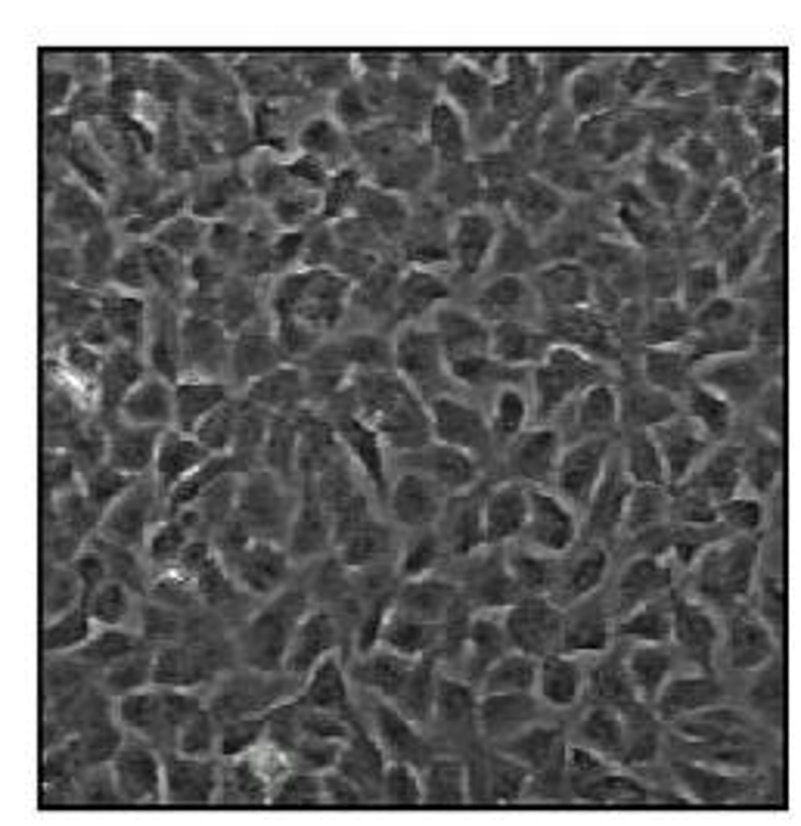
The NB69 cell line is a human-derived cell line commonly used as a model for studying neuroblastoma, which is a common and aggressive pediatric solid tumor that arises from neural crest cells. The cell line was originally developed from the bone marrow of a child with metastatic neuroblastoma, and has since been widely used as an in vitro model for studying this type of cancer. NB69 cells are adherent cells with a characteristic neuronal-like morphology and the extension of neurite-like processes when cultured. The cell line has a rapid doubling time under standard culture conditions, but requires a supplemented medium for survival and exponential growth. Morphologically, the cells are small, polygonal to spindle-shaped, and often appear as loose aggregates, which is a common morphology observed in neuroblast-derived tumor cells.
The NB69 cell line maintains multiple neuroblastoma-specific features which include neuronal marker expression such as tyrosine hydroxylase and GD2 ganglioside along with catecholamine synthesis capabilities. These features make the cell line particularly useful for studying tumor biology, drug resistance, and metastatic potential specific to neuroblastoma. Additionally, NB69 cells have been used in studies exploring the efficacy of combination therapies and the role of the tumor microenvironment in cancer progression.
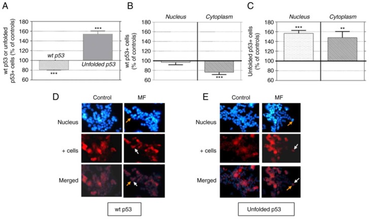
Field exposure to 50 Hz significantly affects wild‑type and unfolded p53 expression in NB69 neuroblastoma cells
Previous studies have shown that intermittent exposure to a 50 Hz, 100 µT sinusoidal magnetic field (MF) promotes proliferation of human neuroblastoma cells, NB69, through a free radical‑dependent activation of the p38 pathway. Martínez et al. investigated whether the oxidative stress‑sensitive protein p53 is a potential target of the MF. To that end, NB69 cells were exposed to short intervals of 30 to 120 min to the aforementioned MF parameters. Immunocytochemical analysis (Fig. 3) showed that MF exposure induced underexpression of wt p53 (18.9±1.3% below controls) and increased the expression of the unfolded isoform (54.1±6.3% above controls). The decrease in wt p53 was cytoplasmic (76.26±2.16% of controls; Fig. 3B), while the increase in unfolded p53 was observed in both the nucleus and cytoplasm (Fig. 3C). Photomicrographs (Fig. 3D and E) illustrated these effects.
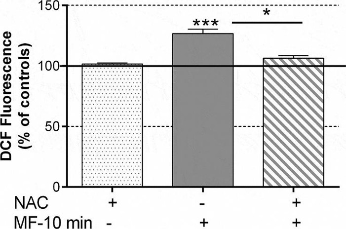
MF Effects on Free Radical Levels of NB69 Cells
Martínez et al. previous studies have shown that intermittent exposure to a 50-Hz, 100-µT sine wave magnetic field (MF) promotes human NB69 cell proliferation through the activation of the epidermal growth factor receptor (EGFR) and MAPK-ERK1/2 and p38 pathways. The present work investigates the MF effects on free radical (FR) production and the potential involvement of NADPH oxidase in the MF-induced activation of MAPK pathways. NB69 cells were exposed to MF or sham and treated with DCFH-DA in the presence or absence of the free radical chelating agent NAC. The fluorescent DCF due to intracellular oxidation of DCFH-DA was analyzed by fluorescence spectroscopy and expressed in terms of ROS production. After 10 minutes of MF exposure followed by a 30-minute post-exposure incubation, the intensity of fluorescent DCF and therefore, of FR production, were significantly increased (26.67% ± 5.9% over controls, Figure 2). Such an increase was not observed in samples exposed to the MF in the presence of NAC.
Ask a Question
Write your own review
Description: Subline of GOTO. Protein-free medium adapted. Cell growth is slow.


Description: Established from the adrenal tumor tissue resected after treatment from a 20-month-old boy of European origin with neuroblastoma (stage III) in 1991
Description: established from the bone marrow metastasis of a 1-year-old female patient with adrenal gland neuroblastoma negative

Description: The line is resistant to infection and focus formation by ecotropic MuLV.
Description: Established from the primary adrenal tumor resected prior to treatment from a 3-year-old boy with rapidly progressing Stage III neuroblastoma in 1989; described to lack MYCN (NMYC, N-myc) ...
- Adipose Tissue-Derived Stem Cells
- Human Neurons
- Mouse Probe
- Whole Chromosome Painting Probes
- Hepatic Cells
- Renal Cells
- In Vitro ADME Kits
- Tissue Microarray
- Tissue Blocks
- Tissue Sections
- FFPE Cell Pellet
- Probe
- Centromere Probes
- Telomere Probes
- Satellite Enumeration Probes
- Subtelomere Specific Probes
- Bacterial Probes
- ISH/FISH Probes
- Exosome Isolation Kit
- Human Adult Stem Cells
- Mouse Stem Cells
- iPSCs
- Mouse Embryonic Stem Cells
- iPSC Differentiation Kits
- Mesenchymal Stem Cells
- Immortalized Human Cells
- Immortalized Murine Cells
- Cell Immortalization Kit
- Adipose Cells
- Cardiac Cells
- Dermal Cells
- Epidermal Cells
- Peripheral Blood Mononuclear Cells
- Umbilical Cord Cells
- Monkey Primary Cells
- Mouse Primary Cells
- Breast Tumor Cells
- Colorectal Tumor Cells
- Esophageal Tumor Cells
- Lung Tumor Cells
- Leukemia/Lymphoma/Myeloma Cells
- Ovarian Tumor Cells
- Pancreatic Tumor Cells
- Mouse Tumor Cells