
HECV
Cat.No.: CSC-6260W
Species: Homo sapiens (Human)
Source: Umbilical Cord
Morphology: continuous culture, grown as monolayer, morphology endothelial
- Specification
- Background
- Scientific Data
- Q & A
- Customer Review
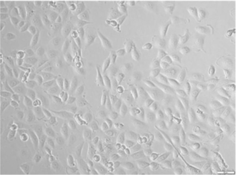
Human Endothelial Cell Line HECV is a continuously growing vascular endothelial cell line from normal human umbilical vein, which was immortalized by stable transfection with SV40 large‑T antigen. This cell line grows as adherent monolayers in routine DMEM medium with 10 % fetal bovine serum, at 37 °C in 5 % CO₂ in air and forms a characteristic cobblestone monolayer with a population‑doubling time of 24-48 h, retaining viability after over 20 passages. The HECV cells express endothelial‑cell markers including CD31, VE‑cadherin and von Willebrand factor that are used to validate the endothelial phenotype.
The HECV cell line effectively builds tubular structures resembling capillaries on Matrigel while responding to VEGF and exhibiting measurable TER which supports its use in barrier‑function studies. The cell line also responds to pro‑inflammatory cytokines (IL‑1β, TNF‑α) with up‑regulation of ICAM‑1/VCAM‑1 and to interleukin‑24 with enhanced migration and modest inhibition of tubulogenesis via AKT/PLCγ signaling. Owing to its stable growth, well‑characterized phenotype and reproducible responses to angiogenic and inflammatory stimuli, HECV is widely used in vascular biology studies, drug screening for anti‑angiogenic agents, endothelial barrier function, and cytokine signaling.
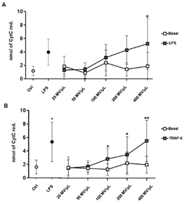
MVs Released by Activated Monocytes and Platelets Induce Oxidative Stress in Endothelial Cells
Circulating microvesicles (MVs) have been implicated in endothelial dysfunction, but no study has directly compared platelet- and monocyte-derived MVs. Brambilla et al. aim to assess their involvement in vessel damage processes, including oxidative stress, inflammation, and leukocyte-endothelial adhesion, by evaluating their effects on human vascular endothelial cells (hECV).
Superoxide anion production was measured in endothelial cells exposed to monocyte- (Mo-) and platelet-derived (Plt-) MVs from resting and activated cells. Results show that MVs from resting cells (25-400 MVs/µL) did not affect superoxide production in human vascular endothelial cells (hECV) (Fig. 1A, B). However, LPS-induced Mo-MVs and TRAP-6-induced Plt-MVs significantly increased superoxide production in a concentration-dependent manner, similar to LPS treatment (Fig. 1A, B). This suggests that MVs from resting cells do not alter endothelial redox balance, while those from activated cells have pro-oxidative effects.
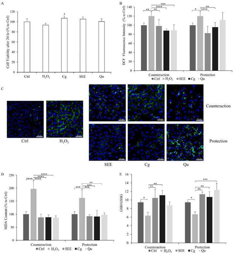
Antioxidant Activity of SEE on Dysfunctional Endothelial Cells
Plants are significant sources of bioactive compounds used in functional foods. In the Mediterranean, Sarcopoterium spinosum is traditionally used for weight loss and diabetes treatment. Given the role of inflammation in diseases like cardiovascular conditions, Zbeeb et al. investigated the antioxidant and cytoprotective properties of an ethanolic extract from S. spinosum fruits (SEE) in a cellular model of endothelium dysfunction.
Initially, they tested SEE, single polyphenols (PPs), and H2O2 for cytotoxicity on HECV cells. No cytotoxicity was observed for 30 µM H2O2, 10 µg/mL SEE, or the PPs corilagin (Cg) and quercetin (Qu) after 24 h (Fig. 2A). Notably, Cg slightly increased cell proliferation. As expected, 30 µM H2O2 significantly increased ROS production (+20% vs. control; p ≤ 0.05) (Fig. 2B). Both SEE and PPs significantly reduced ROS levels when administered after (-22% for SEE, -33% for Cg, -22% for Qu) or before (-24% for SEE, -24% for Cg) H2O2 exposure. Qu pre-treatment showed no significant effect. Fluorescence microscopy visualized these changes (Fig. 2C). Lipid peroxidation was assessed by measuring malondialdehyde (MDA) using a TBARS assay (Fig. 2D). H2O2-insulted cells showed a significant increase in MDA (+63% in protection, +97% in counteraction conditions). In the counteraction condition, all compounds significantly reduced MDA (-110% for SEE, -109% for Cg, -111% for Qu). In the protection condition, SEE and PPs prevented MDA increase (-70% for SEE, -72% for Cg, -64% for Qu). Thus, SEE exhibited strong antioxidant effects, with the counteraction condition being more effective in reducing lipid peroxidation.
Ask a Question
Write your own review
- You May Also Need

Description: Uterine adenosquamous carcinoma. Said CEA and CA125 producing. Cell growth is slow.

Description: Established from tumor tissue from a 77-year-old woman with recurrence of endometrial carcinoma (adenomatous, partly papillary, grade G3) in 1990; described as forming heterotransplantable tumors in ...

Description: Glassy cell carcinoma. TA-4, CA125, neuron-specific enolase producing.
Description: The cells possess alpha keratin, well defined junctional complexes, tonofilaments and surface microvilli.

- Adipose Tissue-Derived Stem Cells
- Human Neurons
- Mouse Probe
- Whole Chromosome Painting Probes
- Hepatic Cells
- Renal Cells
- In Vitro ADME Kits
- Tissue Microarray
- Tissue Blocks
- Tissue Sections
- FFPE Cell Pellet
- Probe
- Centromere Probes
- Telomere Probes
- Satellite Enumeration Probes
- Subtelomere Specific Probes
- Bacterial Probes
- ISH/FISH Probes
- Exosome Isolation Kit
- Human Adult Stem Cells
- Mouse Stem Cells
- iPSCs
- Mouse Embryonic Stem Cells
- iPSC Differentiation Kits
- Mesenchymal Stem Cells
- Immortalized Human Cells
- Immortalized Murine Cells
- Cell Immortalization Kit
- Adipose Cells
- Cardiac Cells
- Dermal Cells
- Epidermal Cells
- Peripheral Blood Mononuclear Cells
- Umbilical Cord Cells
- Monkey Primary Cells
- Mouse Primary Cells
- Breast Tumor Cells
- Colorectal Tumor Cells
- Esophageal Tumor Cells
- Lung Tumor Cells
- Leukemia/Lymphoma/Myeloma Cells
- Ovarian Tumor Cells
- Pancreatic Tumor Cells
- Mouse Tumor Cells