
Rabbit Nucleus Pulposus Cells
Cat.No.: CSC-C5148S
Species: Rabbit
Source: Cartilage
- Specification
- Background
- Scientific Data
- Q & A
- Customer Review
Rabbit nucleus pulposus cells from Creative Bioarray are isolated from the rabbit intervertebral disc tissue. The method we use to isolate rabbit nucleus pulposus cells was developed based on a combination of established and our proprietary methods. The rabbit nucleus pulposus cells are characterized by immunofluorescence with antibodies specific to collagen II. Each vial contains 0.5x10^6 cells per ml and is delivered frozen.
Rabbit nucleus pulposus cells are derived from the nucleus pulposus region of the intervertebral disc. The intervertebral disc is divided into three parts: nucleus pulposus, annulus fibrosus, and cartilage endplate. The nucleus pulposus, which is located in the center, is composed of mainly collagen and proteoglycans, serving as a cushion and shock absorber. Nucleus pulposus cells are mainly located in the central region of the annulus fibrosus, which is the softest part of the disc. They mainly contribute to the maintenance of the hydration and mechanical properties of the intervertebral disc in vivo. By synthesizing and secreting proteoglycans and collagen, they constitute the extracellular matrix of the nucleus pulposus, providing cushioning and resisting compression. In addition, they regulate the metabolic activities and inflammatory responses of the disc, and are related to the process of disc degeneration.
Nucleus pulposus cells have been widely used in vitro studies of the effects of oxidative stress and inflammatory factors (such as IL-1β and TNF-α) on cell function. For example, it was found that nitric oxide (NO) can promote disc degeneration by inhibiting the function of mitochondria, and niacinamide can protect nucleus pulposus cells from damage by inhibiting NO production. Moreover, rabbit nucleus pulposus cells can be used to construct artificial nucleus pulposus tissue for the treatment of spinal degenerative diseases. It was found that rabbit nucleus pulposus cells can be used to grow tissue on scaffold materials through three-dimensional culture techniques, which can grow tissue that is similar to the natural nucleus pulposus, with good mechanical properties and biological activity.
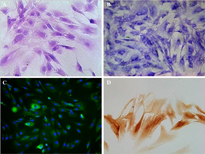 Fig. 1. Staining assessment of NPCs. (a) Haematoxylin–eosin staining. (b) Toluidine blue staining. (c) Immunofluorescence staining. (d) Immunohistochemical staining (Guo MB, Wang DC, et al., 2018).
Lupeol Against High-Glucose-Induced Apoptosis Via Enhancing the Anti-Oxidative Stress in Rabbit Nucleus Pulposus Cells
IDD-induced secondary spinal instability and spinal stenosis are the main causes of low back pain. The reduced number and function of nucleus pulposus cells (NPCs) are the major reasons for IDD. Lupeol, a triterpenoid substance in fruits, vegetables, and Chinese herbal medicines, has been reported to have anti-oxidative, anti-inflammatory, antibacterial, and other benefits in several animal experiments.
Guo's team hypothesized that lupeol can reduce ROS production and inhibit apoptosis of NPCs in a high-glucose environment by antagonizing oxidative stress. The CCK-8 assay showed that cell viability was reduced by concentrations of lupeol more than 60 μg/mL (P < 0.01, Fig. 1a) and had no cytotoxicity by concentrations less than 60 μg/mL. Thus, 60 μg/mL of lupeol was selected for the following experiments. The assay showed no significant difference in cell proliferation activity between the LUP and CON groups, but the HG group had significantly lower activity (P < 0.01). The HG + LUP group had increased activity compared to the HG group (P < 0.05) (Fig. 1b). ROS levels were not significantly different between the LUP and CON groups, but were significantly higher in the HG group (P < 0.01) and lower in the HG + LUP group compared to the HG group (P < 0.05) (Fig. 1c). The study concluded that hyperglycaemia inhibits NPC growth and leads to IDD. Therefore, it was concluded that hyperglycaemia could significantly inhibit the growth of NPCs and lead to IDD.
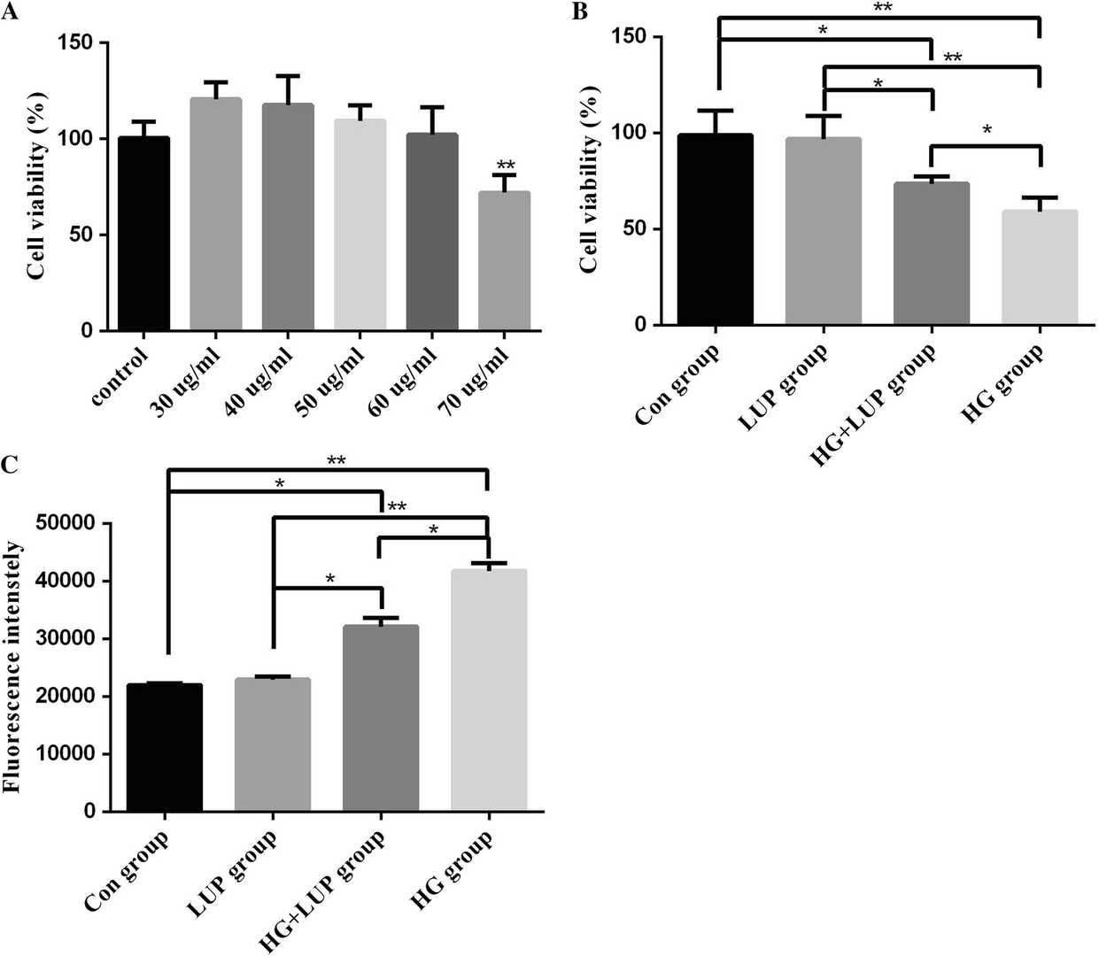 Fig. 1. Lupeol (LUP) protected NPCs from high-glucose-induced cytotoxicity (Guo MB, Wang DC, et al., 2018).
Effects of Hypoxia and ASIC3 on Nucleus Pulposus Cells
Intervertebral disc degeneration, a leading cause of osphyalgia, occurs in an ischemic, hypoxic, acidic environment. This microenvironment, especially acidity, inhibits cell activity and is linked to ASIC3 activation. ASIC3 forms Ca2+-permeable channels at low pH, activating downstream pathways like MAPK and influencing cell behavior. Wang's team explored the effects of hypoxia and acid-sensing ion channel 3 (ASIC3) on nucleus pulposus cells from cell behavior to molecular mechanism.
The mRNA expression of ASIC3 in cells treated with different ASIC3 shRNAs is shown in Figure 2. Three interference sequences for ASIC3 were designed online, with shRNA-ASIC3 2 significantly reducing ASIC3 mRNA expression (P < 0.05), making it suitable for further experiments. Figure 2B demonstrates that the empty vector and shRNA-NC resulted in similar ASIC3 levels as control, whereas shRNA-ASIC3 reduced and ASIC3 expression vector increased ASIC3 expression significantly (P < 0.05), confirming the effectiveness of these interventions. Figure 3 illustrates cell cycle, viability, and apoptosis in various groups. As seen in Figure 3A, 2% O2 treatment increased G1 phase and decreased S phase cell numbers compared to control, with ASIC3+2% O2 showing the most G1 and least S phase cells (P < 0.05). shRNA-ASIC3 relieved G1 phase blockage caused by ASIC3 expression and hypoxia. Figure 3B shows that 48-hour 2% O2 treatment significantly inhibited cell viability (P < 0.05), and Figure 3C reveals increased apoptosis with 2% O2 treatment compared to control (P < 0.05). ASIC3 expression and hypoxia elevated apoptosis, while shRNA-ASIC3 reduced it.
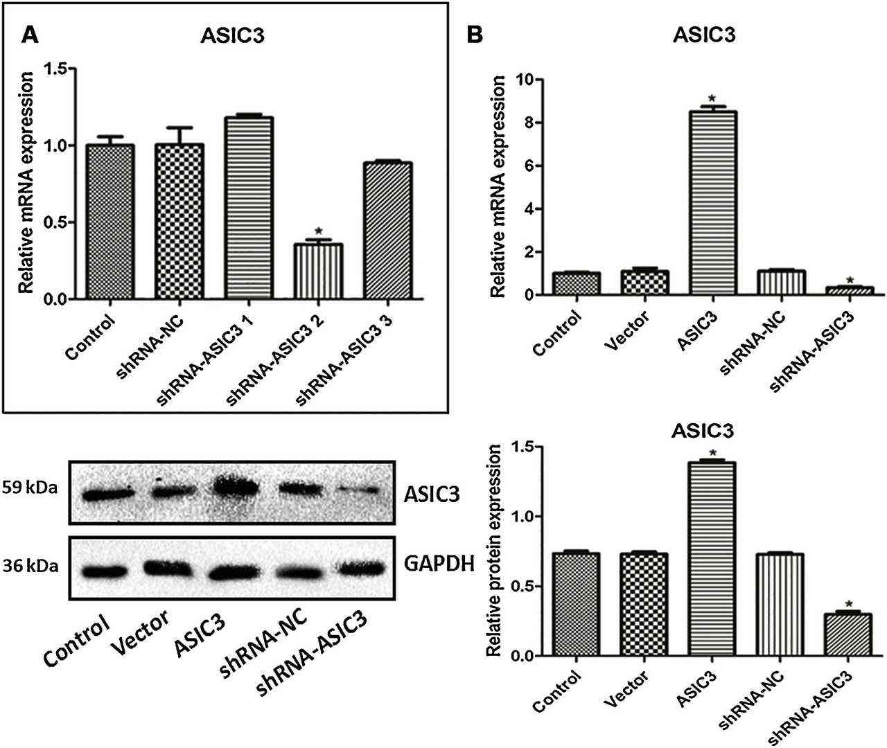 Fig. 2. The expression of ASIC3 in primary rabbit nucleus pulposus cells treated with different ASIC3 shRNA (A) and different ASIC3 expression vectors and interference sequences (B) (Wang D, Zhu H, et al., 2019).
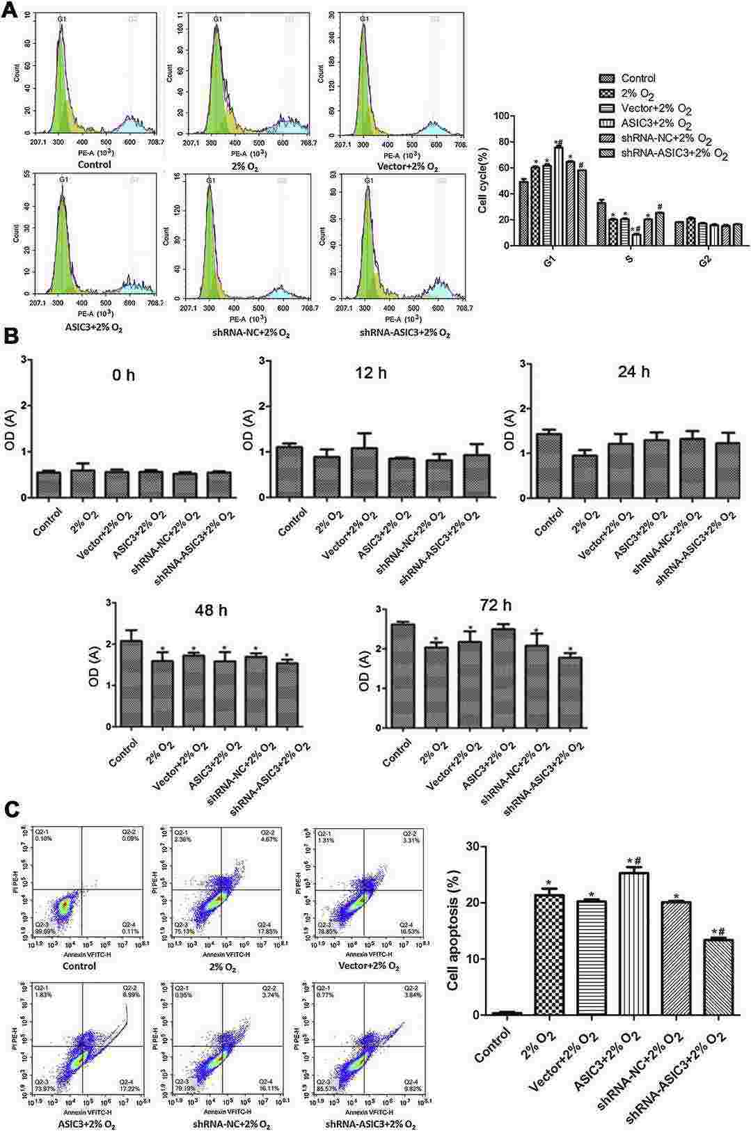 Fig. 3. The cycle (A), viability (B) and apoptosis (C) of primary rabbit nucleus pulposus cells in various groups which were evaluated by flow cytometry and CCK-8 assay (Wang D, Zhu H, et al., 2019).
Ask a Question
Write your own review
Description: Rabbit Hepatocytes are derived from the liver of New Zealand White Rabbit.
Description: The aortic arch is the top part of the main artery carrying blood away from the heart. It is the connection between the ascending and descending aorta, and its central part is formed by the left 4th ...
Description: The synovium secretes synovial fluid, which plays an important role in joint movement. The normal synovium has two layers, a thin cellular layer (luminal layer) and a vascular layer (subintima). ...
Description: The pituitary gland is an important endocrine gland in the body that secretes growth hormone and adrenocorticotropic hormone. It plays an important role in the growth and development of the body, ...
Description: The oral epthelial cells are responsible for important functions, like the primary protection of oral mucosa against external aggressions building a mechanical barrier against microorganisms, ...
Description: The carotid arteries are major blood vessels in the neck that supply blood to the brain, neck, and face. There are two carotid arteries, one on the right and one on the left. In the neck, each ...