ATDC5
Cat.No.: CSC-C6480J
Species: Mus musculus (Mouse)
Morphology: epithelial-like
Culture Properties: Adherent cells
- Specification
- Background
- Scientific Data
- Q & A
- Customer Review
Store in liquid nitrogen.
The ATDC5 cell line is a widely employed murine cellular model derived from a differentiating culture of mouse embryonal carcinoma AT805 cells. It is not a cancer cell line but rather a multipotent mesenchymal progenitor cell line that exhibits a stable and reproducible capacity for chondrogenic differentiation. Since its establishment, ATDC5 has become a cornerstone in vitro system for studying the molecular and cellular events governing chondrocyte differentiation, endochondral ossification, and growth plate development.
ATDC5 cells in a proliferative state exhibit a fibroblast-like morphology. Their defining characteristic is the ability to undergo a well-defined, sequential differentiation program upon stimulation with specific factors, most notably insulin or insulin-like growth factor 1 (IGF-1). This program recapitulates key stages of chondrocyte maturation: 1) Proliferation Phase: Cells proliferate and form dense, nodular aggregates. 2) Differentiation Phase: Cells within the nodules differentiate into chondrocytes, initiating the robust synthesis of a cartilage-specific extracellular matrix (ECM) rich in type II collagen (Col2a1) and aggrecan. 3) Hypertrophic Phase: Following confluence, the culture progresses to a hypertrophic stage, marked by the expression of type X collagen (Col10a1) and alkaline phosphatase (ALP), mimicking the terminal differentiation of growth plate chondrocytes prior to vascular invasion and bone formation.
ATDC5 provides a homogeneous, clonal population that undergoes a predictable and stage-specific differentiation timeline. This allows for precise temporal analysis of gene expression, signaling pathway activation, and ECM deposition during cartilage formation. ATDC5 is extensively used to simulate endochondral bone formation and to study the pathogenesis of skeletal disorders. By subjecting differentiated ATDC5 cultures to pro-inflammatory cytokines (e.g., IL-1β, TNF-α), scientists can mimic aspects of cartilage degradation seen in osteoarthritis (OA), studying the upregulation of matrix-degrading enzymes (MMP-13, ADAMTS-5) and the loss of cartilaginous markers.
FHL2 Deteriorates IL-1β Induced Inflammation, Apoptosis, and Extracellular Matrix Degradation in Chondrocyte-Like ATDC5 Cells
The role of nuclear translocation in osteoarthritis (OA) pathogenesis has garnered increasing attention in recent years. Extensive research has demonstrated that FHL2 acts as a nuclear transmitter through interactions with other nuclear transcription factors. This study aimed to investigate the role of FHL2 in an osteoarthritis cell model.
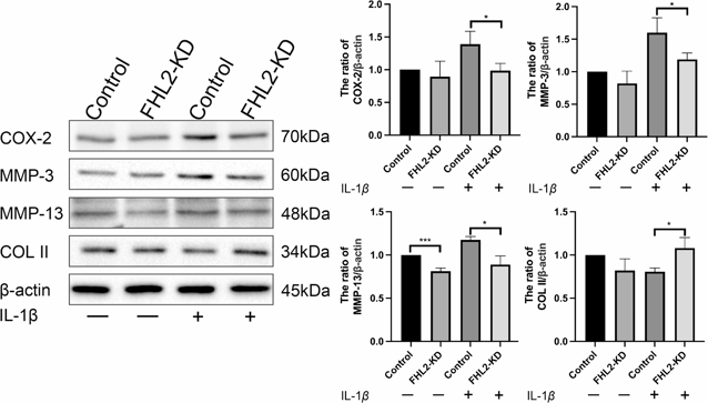
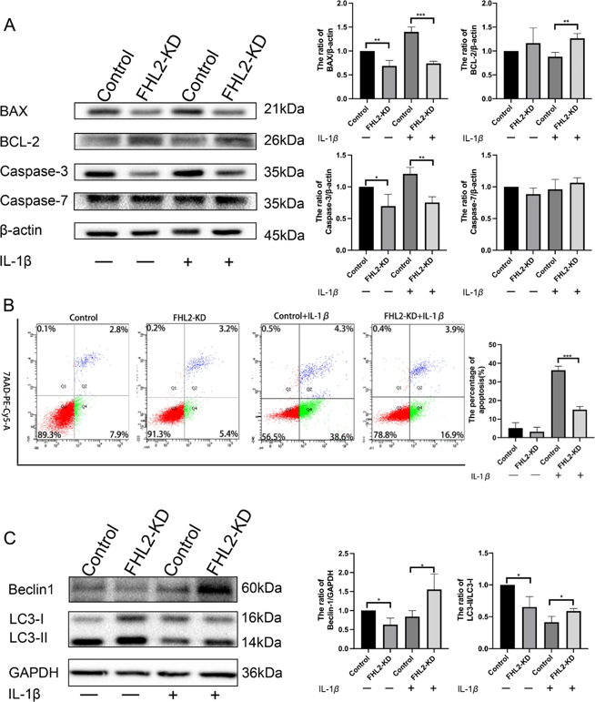
OA cartilage model was established by chondrocyte-like ATDC5 cells induced by 1% insulin-transferrin-selenium and then treated with interleukin-1β (IL-1β, 10 ng/mL). Lentivirus transfection was employed to suppress the expression of FHL2.
The elevated expression of FHL2 occurred in both the cytoplasm and the nucleus. Knockdown of FHL2 could inhibit IL-1β-induced phosphorylation of NF-ĸB p65 and stabilize the extracellular matrix (ECM) by decreasing MMP-3 and MMP-13 expression, to suppress COL II degradation in chondrocyte-like ATDC5 cells. Meanwhile, the knockdown of FHL2-activated autophagy in IL-1β-treated chondrocytes through mTOR signaling, characterized by an increased LC3-II/LC3-I ratio and Beclin-1. FHL2 downregulation inhibited IL-1β-induced apoptosis by suppressing BAX and Caspase-3 expression, while enhancing BCL-2 protein levels. This mechanism may involve AKT phosphorylation and decreased expression of p-NF-ĸB p65.
Ask a Question
Write your own review
- You May Also Need

Description: Described as secreting a mouse monoclonal antibody (IgG2a) detecting all fibers in skeletal muscle and myosin heavy chains on Western blots and detecting mammalian, chicken, zebrafish, axolotl, ...

Description: Animals were immunized with the B6.1 mouse cytotoxic T cell line.


Description: neuroglial and neuronal character coexpressing ependymoma cell line.

Description: Established by irradiation of the adherent cells in long-term bone marrow cultures derived from C3H/HeNSlc strain mice
- Adipose Tissue-Derived Stem Cells
- Human Neurons
- Mouse Probe
- Whole Chromosome Painting Probes
- Hepatic Cells
- Renal Cells
- In Vitro ADME Kits
- Tissue Microarray
- Tissue Blocks
- Tissue Sections
- FFPE Cell Pellet
- Probe
- Centromere Probes
- Telomere Probes
- Satellite Enumeration Probes
- Subtelomere Specific Probes
- Bacterial Probes
- ISH/FISH Probes
- Exosome Isolation Kit
- Human Adult Stem Cells
- Mouse Stem Cells
- iPSCs
- Mouse Embryonic Stem Cells
- iPSC Differentiation Kits
- Mesenchymal Stem Cells
- Immortalized Human Cells
- Immortalized Murine Cells
- Cell Immortalization Kit
- Adipose Cells
- Cardiac Cells
- Dermal Cells
- Epidermal Cells
- Peripheral Blood Mononuclear Cells
- Umbilical Cord Cells
- Monkey Primary Cells
- Mouse Primary Cells
- Breast Tumor Cells
- Colorectal Tumor Cells
- Esophageal Tumor Cells
- Lung Tumor Cells
- Leukemia/Lymphoma/Myeloma Cells
- Ovarian Tumor Cells
- Pancreatic Tumor Cells
- Mouse Tumor Cells