
KYSE-450
Cat.No.: CSC-C0435
Species: Homo sapiens (Human)
Source: Esophagus
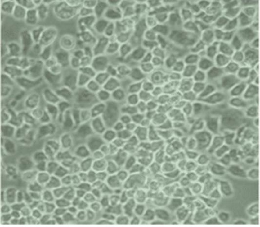
Morphology: epitheloid cells growing adherently as monolayers
Culture Properties: monolayer
- Specification
- Background
- Scientific Data
- Q & A
- Customer Review
Immunology: cytokeratin +, cytokeratin-7 -, cytokeratin-8 +, cytokeratin-17 +, cytokeratin-18 +, cy
KYSE-450 is an immortalized human esophageal squamous cell carcinoma (ESCC) cell line that was isolated in 1990 from a poorly differentiated thoracic ESCC lesion of a male patient. Morphologically, KYSE-450 cells have an epithelial-like morphology, with polygonal or irregular shapes, clear cell borders, high nuclear to cytoplasmic ratio, and prominent nucleoli - typical features of malignant epithelial cells. The cells are grown as adherent monolayers in RPMI 1640 medium supplemented with 10% fetal bovine serum (FBS) and 1% penicillin-streptomycin. Standard culture conditions (37°C, 5% CO₂) produce doubling times of 36-48 hours for the cell population while proper maintenance allows stable growth for more than 50 passages.
Functionally, KYSE-450 cells express typical molecular markers associated with ESCC (e.g., cytokeratins 14 and 19) and contain genomic mutations commonly observed in ESCC (e.g., TP53 mutation, CDKN2A deletion) that underlie their malignant phenotypes, such as uncontrolled proliferation and invasive behavior. As a result, KYSE-450 cells are broadly utilized in studies investigating the pathogenesis of ESCC, for screening of chemotherapeutic (e.g., cisplatin, 5-fluorouracil) and targeted therapeutics and mechanisms of resistance, and for in vitro and in vivo (xenograft) studies of ESCC metastasis and tumor-stroma interactions.
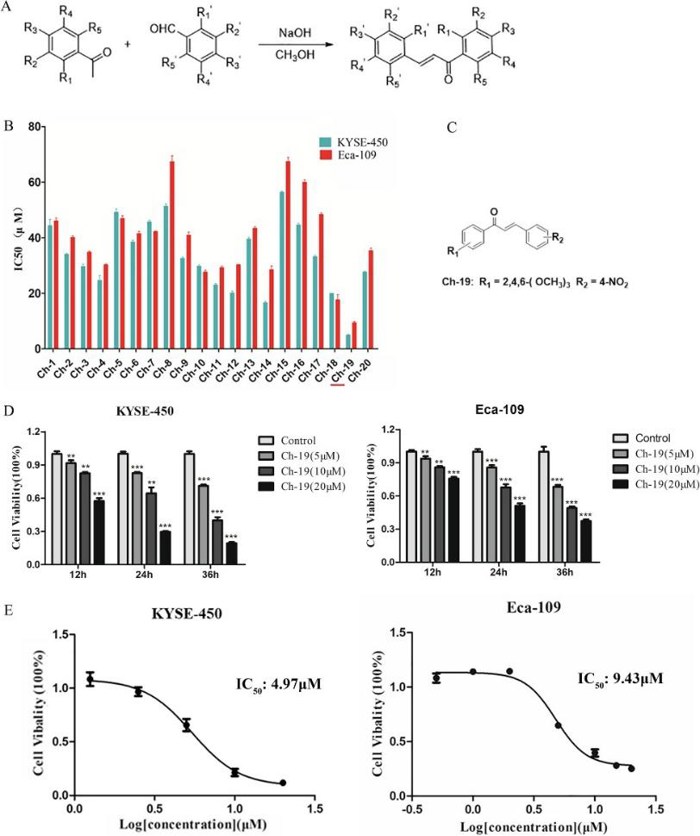
Novel Chalcone Analog Ch-19 Effectively Inhibited ESCC Proliferation In Vitro
Esophageal cancer is characterized by a poor prognosis and low five-year survival rate. Chalcone, a flavonoid, has demonstrated anti-tumor properties in various cancers, but its derivatives' effects on esophageal squamous cell carcinoma are underexplored. In this study, Yang's team designed and synthesized novel chalcone analogs, selecting Ch-19 for its potent anti-tumor activity. They assessed Ch-19's effects on KYSE-450 and Eca-109 esophageal cancer cells (ESCC). CCK-8 assay demonstrated that mean IC50 values of the twenty compounds against both ESCC cells varying from 4.97 to 56.39 µM and 9.43 to 67.40, respectively (Fig. 1B). Notably, Ch-19 exerted the strongest anti-tumor activity against ESCC cells (Fig. 1C), which may be attributed to the substitution of the methoxy group of aromatic ketones and the 4-substitution nitro group of aromatic aldehydes. Moreover, the cell viability was significantly reduced after Ch-19 treatment in both ESCC cells in a dose and time-dependent manner (Fig. 1D-E). The mean IC50 values in KYSE-450 and Eca-109 cells were 4.97 µM and 9.43 µM, respectively.
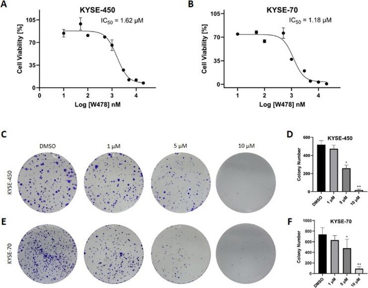
Compound W478 Inhibits the Proliferation of Esophageal Cancer Cell
Esophageal cancer is a serious malignancy with a poor prognosis and high mortality rate. Serine hydroxymethyltransferase 2 (SHMT2) has emerged as a promising target for esophageal cancer therapy. Akram et al. investigated the potential of compound W478, a novel SHMT2 inhibitor, to serve as a lead compound for developing treatments for esophageal carcinoma.
The antiproliferative activity of compound W478 on esophageal cancer cells was assessed using the CCK-8 assay. Results showed that W478 significantly inhibited cell proliferation, with IC50 values of 1.62 μM for KYSE-450 and 1.18 μM for KYSE-70 cells (Fig. 2A and B). A colony formation assay further demonstrated that W478 reduced the number and size of colonies in a dose-dependent manner (Fig. 2C-F). The moderate dose (5 μM) significantly decreased colony numbers, while the maximum dose (10 μM) nearly halted cell growth. These findings indicate that W478 suppresses the clonogenic capacity of KYSE-450 and KYSE-70 cells, supporting its potential as a treatment for esophageal cancer. Additionally, W478's cytotoxicity was tested against HET-1 A and BEAS-2B cells, revealing IC50 values of 7.38 μM and 1.36 μM, respectively, indicating poor selectivity between cancer and normal cells.
Ask a Question
Write your own review
- You May Also Need

Description: Human moderately differentiated squamous cell carcinoma cell line established from esophageal cancer.
Description: Human moderately differentiated squamous cell carcinoma cell line established from esophageal cancer.


Description: Human moderately differentiated squamous cell carcinoma cell line established from esophageal cancer.
Description: Human squamous cell carcinoma from oral cavity via mouse transplantation.

Description: Human squamous cell carcinoma cell line established from recurrent cancer (esophageal cancer).
- Adipose Tissue-Derived Stem Cells
- Human Neurons
- Mouse Probe
- Whole Chromosome Painting Probes
- Hepatic Cells
- Renal Cells
- In Vitro ADME Kits
- Tissue Microarray
- Tissue Blocks
- Tissue Sections
- FFPE Cell Pellet
- Probe
- Centromere Probes
- Telomere Probes
- Satellite Enumeration Probes
- Subtelomere Specific Probes
- Bacterial Probes
- ISH/FISH Probes
- Exosome Isolation Kit
- Human Adult Stem Cells
- Mouse Stem Cells
- iPSCs
- Mouse Embryonic Stem Cells
- iPSC Differentiation Kits
- Mesenchymal Stem Cells
- Immortalized Human Cells
- Immortalized Murine Cells
- Cell Immortalization Kit
- Adipose Cells
- Cardiac Cells
- Dermal Cells
- Epidermal Cells
- Peripheral Blood Mononuclear Cells
- Umbilical Cord Cells
- Monkey Primary Cells
- Mouse Primary Cells
- Breast Tumor Cells
- Colorectal Tumor Cells
- Esophageal Tumor Cells
- Lung Tumor Cells
- Leukemia/Lymphoma/Myeloma Cells
- Ovarian Tumor Cells
- Pancreatic Tumor Cells
- Mouse Tumor Cells