
EOC 20
Cat.No.: CSC-C9179W
Species: Mus musculus (Mouse)
Source: Brain
Morphology: macrophage
- Specification
- Background
- Scientific Data
- Q & A
- Customer Review
EOC‑20 is a murine microglial cell line that was developed in the mid‑1990s from LADMAC‑conditioned medium. It requires CSF‑1 (also known as colony‑stimulating factor 1) for growth and proliferation, which is provided by LADMAC‑conditioned medium. Cells show a semi‑adherent morphology with round to oval somata of approximately 10-12 µm diameter and a relatively large nucleus; they form loose clusters. Cells are typically maintained in DMEM with 10 % fetal bovine serum and 20 % L‑292 conditioned medium, at 37 °C in a humidified 5 % CO₂ incubator; under these conditions, the population doubling time is approximately 24-30 hours, with routine passaging at a 1:2-1:3 ratio every 2-3 days. EOC‑20 cells express typical microglial markers, including CD11b, F4/80, I‑ba1, and MHC II, which is up‑regulated following stimulation with IFN‑γ.
These cells also respond to inflammatory stimuli (LPS, TNF‑α, IFN‑γ) by producing nitric oxide, reactive oxygen species, and pro‑inflammatory cytokines such as IL‑1β and TNF‑α. EOC‑20 cells retain strong phagocytic capacity as determined by fluorescent bead uptake assays. For these reasons, EOC‑20 cells have been used extensively to investigate microglial signaling cascades (e.g., NF‑κB, MAPK, JAK/STAT), screen for anti‑inflammatory or neuroprotective agents, and model aspects of neurodegenerative diseases, such as Alzheimer's disease and Parkinson's disease. In addition, this line is highly amenable to genetic manipulation, allowing detailed studies of key genes involved in microglial activation, including TLR4, NLRP3, and CX3CR1.
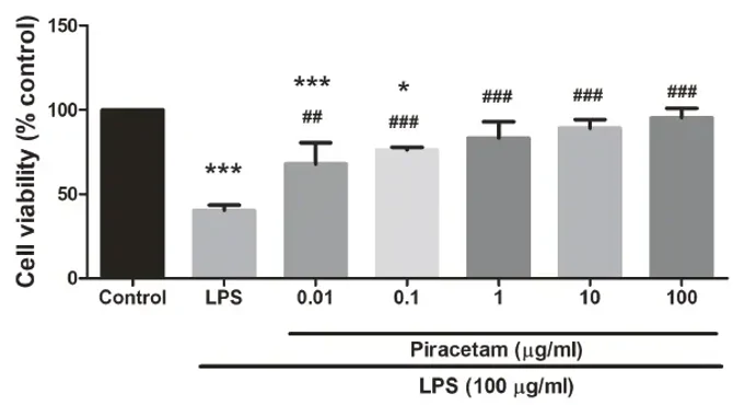
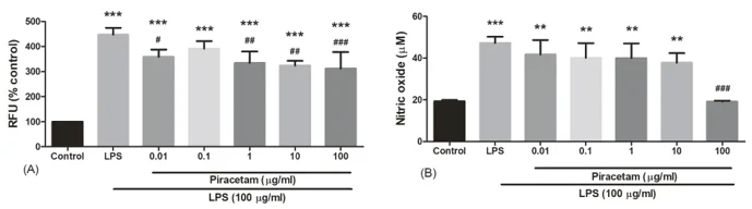
Pre-Treatment with Piracetam Reduced the Level of Intracellular ROS and NO against LPS-Induced Oxidative Stress in EOC-20 Cells
Piracetam is a popular nootropic. Mani et al. investigated its neuroprotective activity against LPS-induced toxicity. Mani used EOC-20 microglial cells to measure ROS and NO production in vitro after LPS treatment and piracetam pre-treatment.
EOC-20 cells treated with 100 µg/ml LPS for 24 h showed 60% cell death by MTT assay. Piracetam (0.01-100 µg/ml) pre-treatment for 24 h significantly improved cell survival with viability reaching 68%-95%. The highest dose of piracetam (100 µg/ml) was the most protective against LPS-induced cell death in a dose-dependent manner (Fig. 1). Figure 2 demonstrates the effect of piracetam on LPS-induced ROS and NO in EOC-20 cells. LPS treatment significantly increased intracellular ROS and NO levels. Piracetam (0.01-100 µg/ml) pre-treatment significantly decreased LPS-induced ROS and the highest piracetam dose (100 µg/ml) significantly reduced NO. Piracetam significantly protected against LPS-induced oxidative stress in EOC-20 cells.
Ask a Question
Write your own review
- You May Also Need

Description: Described as secreting a mouse monoclonal antibody (IgG2a) detecting all fibers in skeletal muscle and myosin heavy chains on Western blots and detecting mammalian, chicken, zebrafish, axolotl, ...

Description: Animals were immunized with the B6.1 mouse cytotoxic T cell line.


Description: neuroglial and neuronal character coexpressing ependymoma cell line.

Description: Established by irradiation of the adherent cells in long-term bone marrow cultures derived from C3H/HeNSlc strain mice
- Adipose Tissue-Derived Stem Cells
- Human Neurons
- Mouse Probe
- Whole Chromosome Painting Probes
- Hepatic Cells
- Renal Cells
- In Vitro ADME Kits
- Tissue Microarray
- Tissue Blocks
- Tissue Sections
- FFPE Cell Pellet
- Probe
- Centromere Probes
- Telomere Probes
- Satellite Enumeration Probes
- Subtelomere Specific Probes
- Bacterial Probes
- ISH/FISH Probes
- Exosome Isolation Kit
- Human Adult Stem Cells
- Mouse Stem Cells
- iPSCs
- Mouse Embryonic Stem Cells
- iPSC Differentiation Kits
- Mesenchymal Stem Cells
- Immortalized Human Cells
- Immortalized Murine Cells
- Cell Immortalization Kit
- Adipose Cells
- Cardiac Cells
- Dermal Cells
- Epidermal Cells
- Peripheral Blood Mononuclear Cells
- Umbilical Cord Cells
- Monkey Primary Cells
- Mouse Primary Cells
- Breast Tumor Cells
- Colorectal Tumor Cells
- Esophageal Tumor Cells
- Lung Tumor Cells
- Leukemia/Lymphoma/Myeloma Cells
- Ovarian Tumor Cells
- Pancreatic Tumor Cells
- Mouse Tumor Cells