
CAL-72
Cat.No.: CSC-C0473
Species: Homo sapiens (Human)
Source: Bone
Morphology: fibroblastoid cells growing adherently in monolayers
Culture Properties: monolayer
- Specification
- Background
- Scientific Data
- Q & A
- Customer Review
Immunology: cytokeratin -, desmin -, endothel -, GFAP -, HMB-45 -, neurofilament -, vimentin +
Viruses: PCR: EBV -, HBV -, HCV -, HIV -, HTLV-I/II -, SMRV -
The CAL-72 cell line originates from an osteosarcoma patient and represents a primary osteosarcoma cancer cell line. Osteosarcoma represents a highly malignant bone tumor which usually develops in the epiphysis or diaphysis regions of long bones including the distal femur and proximal tibia. The CAL-72 cell line displays osteoblast-like morphology together with pronounced abilities to proliferate rapidly and migrate effectively. When exposed to particular conditions these cells develop into sarcospheres, which represent a three-dimensional arrangement of osteosarcoma cells helping researchers study their growth patterns and metastatic capabilities. The cultivation of CAL-72 cells reveals substantial mitochondrial activity which appears to connect with their energy requirements and metabolic functions.
The CAL-72 cell line serves as a crucial model for researching osteosarcoma pathogenesis while examining drug sensitivity and resistance mechanisms. Research has shown that CAL-72 cells demonstrate extreme sensitivity to specific kinase inhibitors including Torin2 and Ganetespib which helps guide new treatment approaches. The sensitivity of the CAL-72 cell line to multiple drugs makes it a well model for testing anticancer drug effectiveness.
CAL-72 and Saos-2 Human Osteosarcoma Cell Lines Formed Sarcospheres
Osteosarcoma is a challenging cancer with poor prognosis due to its aggressive nature and frequent lung metastasis. Although treatment improvements have been made, long-term survival rates remain unchanged, necessitating new approaches. Stem cells, known for self-renewal and pluripotency, have been linked to various cancers. Lim et al.uses osteosarcoma cell lines under non-adherent conditions to culture sarcospheres, enabling the identification and characterization of cancer stem cells.
CAL-72 and SaOs-2 human osteosarcoma cell lines formed sarcospheres within 7 days of cultivation (Fig. 1). About 1.5% of CAL-72 and 1.0% of SaOs-2 cells formed sarcospheres, whereas human bone marrow-derived mesenchymal stem cells did not survive for over a week in non-adhesive conditions without FBS. Sarcospheres from osteosarcoma cell lines developed into cancer cells under adhesive conditions but remained sarcospheres in non-adhesive conditions (Fig. 2). Despite repeated attempts, sarcospheres and monolayer cancer cells did not grow beyond their original states in non-adhesive conditions. Under normal monolayer conditions, sarcospheres grew at similar rates across generations, achieving sufficient cell concentration by the 7th generation similar to the 1st.
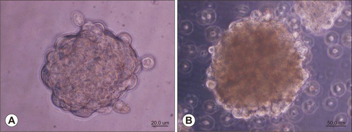 Fig. 1. Phase contrast images of sarcospheres from human osteosarcoma cell lines (ALP stainng) (Lim C, Roh YH, et al., 2022).
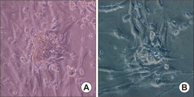 Fig. 2. Adherent cancer cells expanded from the spheres when sarcosphere reattached to adherent plate after removal from the non-adherent culture (ALP stainng) (Lim C, Roh YH, et al., 2022).
MO-OH-Nap Induces Apoptosis in OS Cells
Osteosarcoma (OS) is a challenging bone cancer with limited treatment progress, underscored by low survival rates for metastatic cases. Tropolones, small aromatic compounds from plants, have shown antiproliferative effects in cancer, targeted here due to their iron-chelating properties in cancer therapy. Haney et al. aims to explore the effects of a novel tropolone, MO-OH-Nap, on human OS cell lines, hypothesizing that it induces apoptosis and affects cellular iron dynamics.
They assessed MO-OH-Nap's effects on five human OS cell lines (143B, CAL-72, HOS, MG-63, SaOS-2) using MTT assays. A 72-hour incubation showed concentration-dependent cytotoxicity across all lines, with HOS being most sensitive (IC50 = 0.67 μM) and SaOS-2 the least (IC50 = 5.93 μM; Fig. 3b). Similarly, 48-hour incubation yielded concentration-dependent cytotoxicity. Flow cytometry confirmed that MO-OH-Nap induces early and late apoptosis at 48 and 72 hours (Fig. 3c). Immunoblot analysis revealed PARP cleavage and caspase accumulation in all lines at both time points (Fig. 3d), with four lines showing these effects after 24-hour exposure to 10 μM MO-OH-Nap. These findings highlight MO-OH-Nap's induction of apoptosis in OS cell lines.
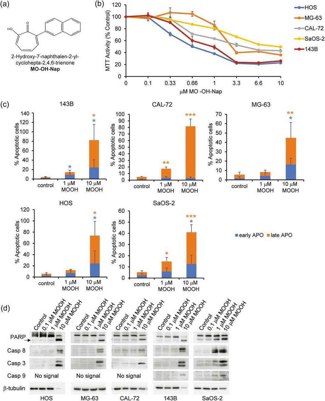 Fig. 3. MO-OH-Nap induces apoptosis in OS cells (Haney S L, Feng D, et al., 2023).
Ask a Question
Write your own review
- You May Also Need

Description: Organism: Homo sapiens (human)Ethnicity: CaucasianAge/Stage: 36 years of ageGender: maleTissue: sarcomaGrowth Properties: monolayerDescription: in vitro established from a primary skin sarcoma (sole ...

Description: Human cell line derived from epithelioid sarcoma. Derived from a different patient from the patient of HS-ES-1 cell line.
Description: Established from an endometrial stromal sarcoma of the uterus of a 76-year-old Caucasian woman; cells were described as demonstrating a mainly mesenchymal phenotype with signs of epithelial ...

Description: Established from the alveololar rhabdomyosarcoma of the liver of a girl after chemotherapy; cells were described to contain a deletion mutation of p53 and to be resistant to FAS- and TRAIL-induced ...

- Adipose Tissue-Derived Stem Cells
- Human Neurons
- Mouse Probe
- Whole Chromosome Painting Probes
- Hepatic Cells
- Renal Cells
- In Vitro ADME Kits
- Tissue Microarray
- Tissue Blocks
- Tissue Sections
- FFPE Cell Pellet
- Probe
- Centromere Probes
- Telomere Probes
- Satellite Enumeration Probes
- Subtelomere Specific Probes
- Bacterial Probes
- ISH/FISH Probes
- Exosome Isolation Kit
- Human Adult Stem Cells
- Mouse Stem Cells
- iPSCs
- Mouse Embryonic Stem Cells
- iPSC Differentiation Kits
- Mesenchymal Stem Cells
- Immortalized Human Cells
- Immortalized Murine Cells
- Cell Immortalization Kit
- Adipose Cells
- Cardiac Cells
- Dermal Cells
- Epidermal Cells
- Peripheral Blood Mononuclear Cells
- Umbilical Cord Cells
- Monkey Primary Cells
- Mouse Primary Cells
- Breast Tumor Cells
- Colorectal Tumor Cells
- Esophageal Tumor Cells
- Lung Tumor Cells
- Leukemia/Lymphoma/Myeloma Cells
- Ovarian Tumor Cells
- Pancreatic Tumor Cells
- Mouse Tumor Cells