- Specification
- Background
- Scientific Data
- Q & A
- Customer Review
Rat Tenocytes from Creative Bioarray are isolated from the tendon tissue of rats. The method we use to isolate renal collecting duct epthelial cells was developed based on a combination of established and our proprietary methods. Rat Tenocytes from Creative Bioarray are characterized by immunofluorescence with antibodies specific to type I collagen. Each vial contains 0.5x10^6 cells per ml and is delivered frozen.
The main source of rat tenocytes are rat tendons particularly those from the patellar and Achilles regions. Cells displaying flat fibroblast morphology serve essential functions in tendon tissue development. Under optimal conditions such as serum-enriched media they exhibit limited proliferation capabilities. High glucose conditions and drugs like lidocaine disrupt the proliferation of these cells. Mechanical stress affects tendon cell growth and differentiation while low-intensity stretching promotes their development into mature tendon cells. Tenocytes mainly produce collagen and other extracellular matrix elements to preserve tendon structure and function. These cells produce collagen-related genes including types I and III collagen and participate in inflammatory responses and tissue repair processes which are essential for tendon health.
Rat tenocytes are vital in regenerative medicine and tissue engineering, with applications in tendon tissue engineering for constructing artificial tendons, drug screening to study effects on tenocyte function, co-culture systems with mesenchymal stem cells for gene expression and differentiation studies, and disease modeling for conditions like gout and tendinitis.
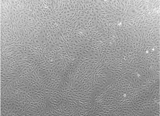 Fig. 1. Micrographs of tenocytes derived from the Achilles tendon in the rat ((Oreff G L, Fenu M, et al., 2021).
In Vitro Tenocyte-Protective Effectiveness of Dehydroepiandrosterone Against High Glucose-Induced Oxidative Stress
Diabetes exacerbates tendinopathy through oxidative stress and ROS production, often via NADPH oxidase (NOX). Mukohara et al. examined DHEA's antioxidant effects on rat tenocytes in low and high glucose conditions and diabetic rat tendons. By analyzing expressions of NOX, IL-6, MMP-2, TIMP-2, and collagen, both in vitro and in vivo, it aims to assess DHEA's effectiveness in reducing oxidative stress and improving tendon matrix turnover.
The WST assay showed rat tenocytes viability improved with 1, 10, and 20 μM DHEA, with 10 μM being the most effective, while 20 and 50 μM reduced viability in a dose-dependent manner (Fig. 1). At 48 hours, NOX1 and IL-6 mRNA levels were higher in the high glucose (HG) DHEA- group than in the low glucose (LG) DHEA- group, but lower in the HG DHEA+ group compared to the HG DHEA- group, with no change in NOX4 mRNA across groups (Fig. 2). ROS-positive cells were stained green, indicating greater ROS in the HG DHEA- group than the LG DHEA- group. ROS was lower in the HG DHEA+ group compared to the HG DHEA- group, with no difference between LG groups (Fig. 3A and B).
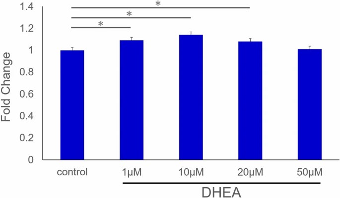 Fig. 1. Cell proliferation (Mukohara S, Mifune Y, et al., 2021).
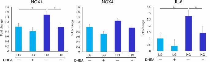 Fig. 2. Quantitative real-time PCR analysis (Mukohara S, Mifune Y, et al., 2021).
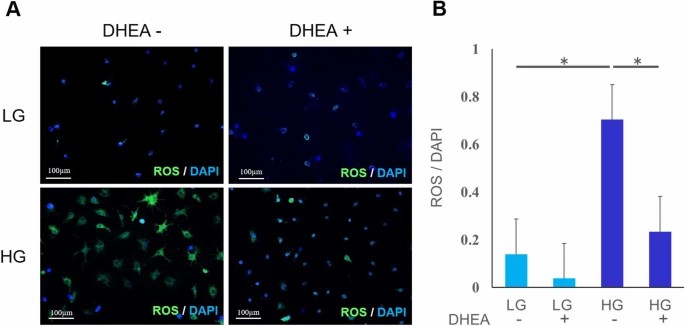 Fig. 3. ROS accumulation (Mukohara S, Mifune Y, et al., 2021).
Comparison of Tenocytes Proliferation, Migration and Gene Expression in Five Animal Species
Tendinopathy, a prevalent musculoskeletal disorder, involves inflammation and impaired healing, costing billions annually. Animal models are essential for studying tendinopathy as human tissue is limited to severe cases. However, species differences affect inflammation responses and pathophysiology. Oreff et al. evaluated cellular and molecular inflammation features in tenocytes from humans and model animals (mouse, rat, sheep, and horse) to guide the selection of suitable models for tendon research.
Proliferation, migration, and gene expression of tenocytes from five species were compared under healthy and inflammatory conditions. Under healthy conditions (Fig. 4), horse tenocytes had the highest proliferation rates, while mouse tenocytes the lowest. Human tenocytes showed low proliferation and couldn't double in 48 hours, with sheep and rat in between. Figure 4 demonstrates significant slope differences among species but low variance within species, with model species significantly differing from humans. Under constant inflammation (10 ng/ml IL1β and TNFα), sheep tenocyte proliferation dropped to human levels, significantly altering their slope difference from the healthy state. Other slope differences under varying inflammatory conditions were not significant.
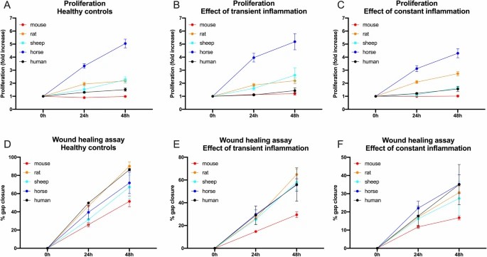 Fig. 4. The proliferation capacity (A–C) of tenocytes from 5 different mammalian species (mouse, rat, sheep, horse, and human) under healthy (Ctr/A), transient (TI/B) and constant (CI/C) inflammatory condition is illustrated as fold increase over the course of 2 days (Oreff G L, Fenu M, et al., 2021).
Ask a Question
Write your own review

Description: The thoracic aorta is located in the chest cavity and gives off arteries that branch to the esophagus, pericardium, lungs, and trachea. The thoracic aorta can be subdivided into the ascending aorta, ...

Description: Rat Podocytes are isolated from normal rat kidney. The cells are characterized by immunofluorescence with antibodies specific to podocin, Ang1, Nephrin, ACTN4, NPHS2. T25 flasks is required for cell ...

Description: Rat Bronchial Smooth Muscle Cells are isolated from normal rat bronchi tissue. Rat Bronchial Smooth Muscle Cells are characterized by immunofluorescence with antibodies specific to alpha-actin. T25 ...

Description: Guinea Pig Endothelial Cells from Creative Bioarray are isolated from guinea pig tissue. Prior to shipping, cells at passage 2 are detached from flasks and immediately cryopreserved in vials. Each ...

Description: Rat Lung Epithelial Cells are isolated from normal rat lung tissue. The cells are characterized by immunofluorescence with antibodies specific to CK-18, CK-19. T25 flasks is required for cell ...

Description: Rat Vein Endothelial Cells from Creative Bioarray are isolated from inferior vena cava tissue of 6-8 week old laboratory Sprague-Dawley rat. Rat Vein Endothelial Cells are grown in T75 tissue culture ...