
Human Pulmonary Vein Endothelial Cells
Cat.No.: CSC-C4866L
Species: Human
Source: Lung; Vein
Cell Type: Endothelial Cell
- Specification
- Background
- Scientific Data
- Q & A
- Customer Review
Never can cryopreserved cells be kept at -20 °C.
Human Pulmonary Vein Endothelial Cells (HPVECs) are human cells that line the interior surface of pulmonary veins, which are blood vessels that are part of the pulmonary circulation carrying oxygenated blood from the lungs to the heart. HPVECs display multiple important characteristics such as preserving vascular endothelium stability and controlling vascular function while facilitating gas exchange and inflammation response, they possess oval or ovoid nuclei and contain Weibel-Palade bodies which regulate vascular operations.
HPVECs have several potential applications in research and medicine, including modeling of pulmonary diseases, drug screening and development, regenerative medicine, and tissue engineering. HPVECs have been used to study various pulmonary diseases, such as pulmonary hypertension, pulmonary fibrosis, and pulmonary embolism, and have been used to model disease mechanisms. HPVECs have also been used for drug screening and the development of drug delivery systems, particularly for anti-inflammatory, anticoagulant, and vascular-protective drugs. Endothelial progenitor cells (EPCs) derived from HPVECs have been explored for their potential use in vascular regeneration and tissue repair, with potential applications in the treatment of cardiovascular and ischemic diseases.
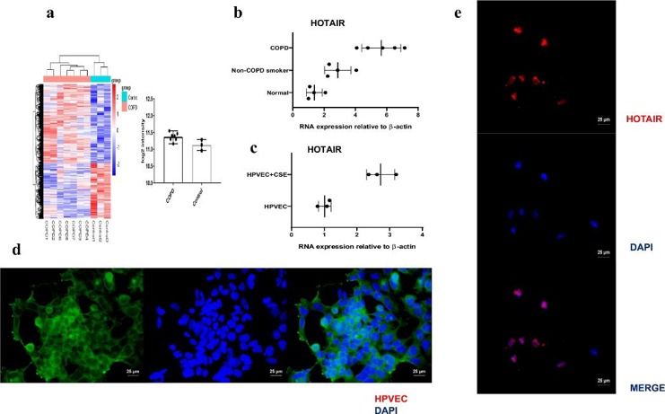
Expression of HOTAIR is Specifically Up-Regulated in COPD Patient Lungs and CSE-Induced HPVEC
COPD is marked by increased HPVEC apoptosis. LncRNAs are being increasingly recognized to participate in many biological processes through diverse mechanisms, such as chromatin modification, protein activity regulation, gene imprinting and so on. Dai et al. investigates the role of the long noncoding RNA HOTAIR in pulmonary vascular endothelial cell (HPVEC) apoptosis and whether HOTAIR promotes HPVEC apoptosis through DNMT1-mediated hypermethylation of the Bcl-2 promoter in COPD.
They collected lung tissues from 13 patients who underwent resection of peripheral solitary pulmonary nodules or lesions. Among them, 4 were normal nonsmokers, 4 were smokers without COPD, and the remaining 5 were smokers with spirometry-defined COPD. An lncRNA array was used to measure differentially expressed lncRNAs in COPD and non-COPD lung tissues. HOTAIR was significantly up-regulated in COPD patients (≥2-fold change) by hierarchical clustering (Fig. 1a). qRT-PCR analysis confirmed overexpression of HOTAIR in COPD smokers, while expression in nonsmokers and non-COPD smokers was very low (Fig. 1b). Similarly, HOTAIR expression was detected in HPVEC and CSE-induced HPVEC, with HPVEC labeled by Recombinant Von Willebrand Factor (VWF) (Fig. 1d). HOTAIR was the most up-regulated lncRNA in CSE-induced HPVEC compared with HPVEC (Fig. 1c). RNA in situ hybridization (RNA-ISH) revealed specific nuclear signals for HOTAIR in HPVEC (Fig. 1e). These results suggest that HOTAIR may play an important role in COPD.
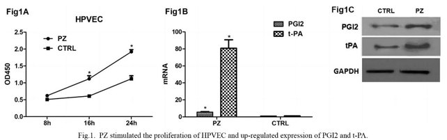
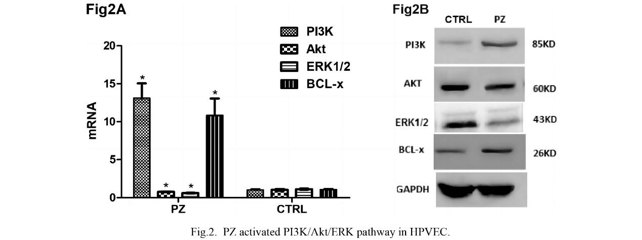
Protein Z Promotes Proliferation in Vascular Endothelial Cell Mediated by Chemokine C-X-C-Motif Receptor 4
Preeclampsia (PE) is associated with changes in vascular endothelial cells. Previous studies have shown that Protein Z (PZ) can promote the proliferation of vascular endothelial cells, though the specific mechanism is unknown. Zhang’s team explored how PZ affects endothelial cell proliferation by investigating its interaction with Chemokine C-X-C-motif receptor 4 (CXCR4) and its impact on the PI3K/Akt/ERK signaling pathway.
Due to the fact that proliferation is essential for angiogenesis and anticoagulation, in the next step we investigated the effect of PZ for the activity of cells, including proliferation, expression of anticoagulation markers PGI2 and t-PA. As shown in Fig. 2A, the results of CCK8 assay indicated that PZ treatment led to increased proliferation of HPVEC. In Fig. 2B and C, the results of qRT-PCR and western blotting shown that the expression of gene and protein of PGI2 and t-PA were up-regulated. It is well known that PI3K/Akt/ERK signaling pathway is critical for the regulation of endothelial cell proliferation. Thus, we examined whether PZ affects this pathway in HPVEC. As shown in Fig. 3A, the results of qRT-PCR revealed the expression of PI3K/Akt/ERK and BCL-x genes were up-regulated by the PZ treatment. In Fig. 3B, the results of western blotting are consistent with qRT-PCR results, showing that the protein level of PI3K/Akt/ERK and BCL-x were promoted by PZ.
Ask a Question
Write your own review
Description: Human Pulmonary Artery Fibroblasts are isolated from normal human pulmonary artery.

Description: Primary Human Fetal Lung Fibroblast Cells were initiated from human lung tissue (normal 25 week gestation).These cells were originated using Complete Serum-Free Medium Kit With SuperFuel™, are ...

Description: GFP-Human Lung Airway Smooth Muscle Cells (GFP-HLAWSMCs) provided by Creative Bioarray are puromycin-selected from primary human lung airway smooth muscle cells infected with lentivirus expressing ...

Description: ACE2-GFP Expressing Human Pulmonary Artery Endothelial Cells (ACE2-GFP-HPAECs) provided by Creative Bioarray are puromycin-selected from Human Pulmonary Artery Endothelial Cells infected with ...
Description: HTSMC from Creative Bioarray Research are isolated from human trachea. HTSMC are cryopreserved at secondary culture and delivered frozen. Each vial contains >5 x 10^5 cells in 1 ml volume. HTSMC are ...

Description: The human type 1 alveolar epithelial cells from Creative Bioarray are isolated from human alveolar tissue. These cells are shipped at passage 2 and maintained in SuperCult® human type 1 alveolar ...