
L-428
Cat.No.: CSC-C0322
Species: Homo sapiens (Human)
Source: Pleural Effusion
Morphology: single cells in suspension or as clusters; polymorph cells, some cells have "spikes", 1-5% giant multinucleated cells
Culture Properties: suspension
- Specification
- Background
- Scientific Data
- Q & A
- Customer Review
- Documents
Immunology: CD2 -, CD3 -, CD13 -, CD14 -, CD15 +, CD19 -, CD25 -, CD30 +, CD34 -
Viruses: ELISA: reverse
The L-428 cell line is a well-established neoplastic cell line derived from the pleural effusion of a female patient with Hodgkin's disease of the nodular sclerosing type. The L-428 cell line exhibits several distinctive characteristics, including aneuploidy and multiple structural and numerical chromosomal abnormalities, which are typical markers of its neoplastic nature. L-428 cells lack surface or cytoplasmic immunoglobulins, despite their derivation from a lymphoid malignancy, which suggests significant differentiation from normal lymphocytes. The absence of Epstein-Barr Virus (EBV) antigens, such as EBNA and VCA, further distinguishes L-428 from other EBV-positive Hodgkin's lymphoma cell lines. These cells also lack lysozyme, peroxidase, and chloroacetate esterase activity, reinforcing their distinction from myeloid cells, monocytes, or macrophages.
L-428 cells closely resemble the Reed-Sternberg (RS) and Hodgkin (H) cells, which are hallmark cells of Hodgkin's lymphoma. These cells have a unique phenotype distinct from typical B cells, T cells, and other hematopoietic cell types, contributing to ongoing debates about the exact cellular origin of RS and H cells. Morphologically, L-428 cells exhibit a range of sizes, from small mononuclear cells to large multinucleated cells, with some cells displaying villous projections on their membranes. The cells are also striking for their large, often kidney-shaped nucleoli. Functionally, L-428 cells express Ia-like antigens and T-cell receptors, but lack other common lymphoid and myeloid markers. This unique immunophenotype, combined with the chromosomal and morphological features, supports the classification of L-428 as a model of Hodgkin's lymphoma, especially for studying the biological properties of RS and H cells.
Mycobacterial Antigen Ag85B Restrains Hodgkin Lymphoma Tumor Growth by Inhibiting Autophagy
The growth of the B-cell lymphoma subtype, Hodgkin lymphoma (HL), is associated with increased autophagy. A mycobacterial antigen, Ag85, has been reported to inhibit cell autophagy under a variety of conditions. Whether Ag85 could inhibit autophagy in HL is unknown.
The human HL cell line, L-428, was cultured and subjected to Ag85B treatment. Autophagy in L-428 cells was evaluated through western blotting analysis, immunohistochemistry, and transmission electron microscopy. Apoptosis in these cells was measured using flow cytometry and western blotting. The associated signaling pathways were also analyzed utilizing western blotting. The results suggest that Ag85B inhibits HL growth via autophagy regulation. Current treatments for HL are associated with adverse events; therefore, Ag85B-mediated autophagy inhibition might be a promising strategy in to treat HL.
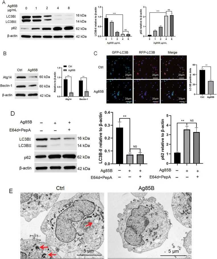 Fig. 1. Ag85B inhibits autophagy in L-428 cells (Cheng, Yongfeng, et al. 2025).
Protein Kinase CK2 Subunits are Unbalanced in Classical Hodgkin Lymphoma, Which May Represent a New Target for Therapy
In classical Hodgkin lymphoma (cHL), tumor cell survival depends on the activation of NF-κB, JAK/STAT and PI3K/Akt signaling pathways. CK2 is a highly conserved serine/threonine kinase, consisting of two catalytic (α) and two regulatory (β) subunits, which is involved in several cellular processes and both subunits were found overexpressed in solid tumors and hematologic malignancies.
This study investigated the expression of CK2α and CK2β in a panel of HL cell lines, analyzed CK2 mediated activation of survival signaling pathways and investigated the capability of inhibiting CK2 with the ATP-competitive inhibitor CX-4945/Silmitasertib along with monomethyl auristatin E (MMAE) to trigger HL cell apoptosis and assess PD-L1 expression levels. These findings present a novel potential target to overcome resistance or to increase MMAE cytotoxicity.
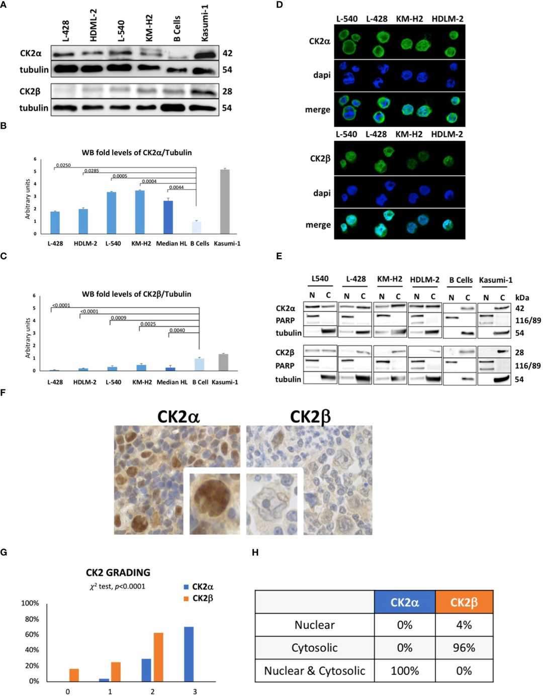 Fig. 2. Protein expression levels of CK2 alpha (α) and beta (β) subunits in HL cell lines (Ruggeri, Edoardo, et al. 2024).
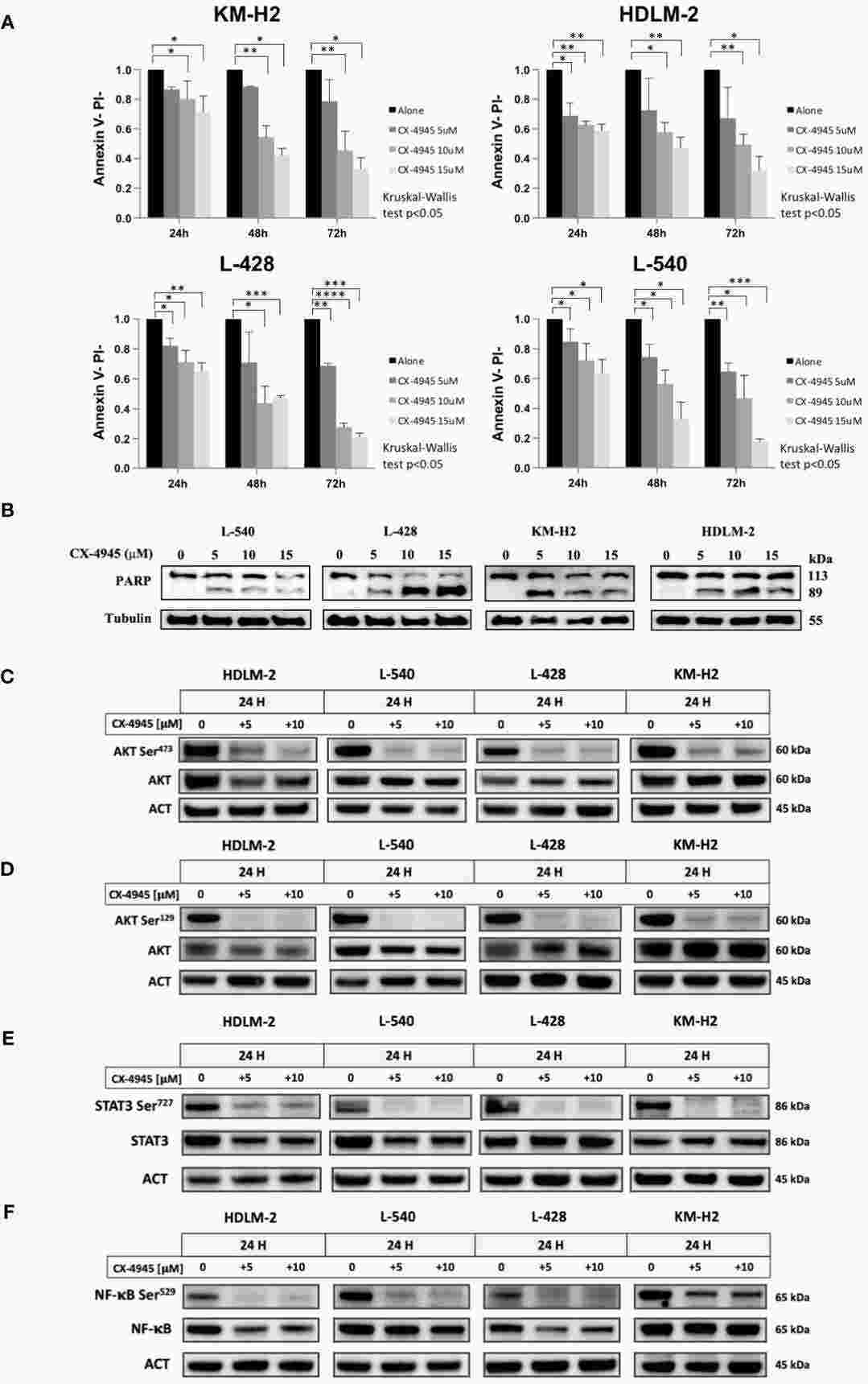 Fig. 3. Apoptotic Effect of CX-4945 in HL Cell Lines (Ruggeri, Edoardo, et al. 2024).
Ask a Question
Write your own review
- You May Also Need

Description: Established in 2007 from the bone marrow mononuclear cells of an 82-year-old Japanese man with diffuse large B-cell lymphoma in the leukemic phase

Description: Established from the bone marrow of a 28-year-old man who developed the terminal leukemic phase of lymphosarcoma in 1976

Description: This cell line was derived from the bone marrow aspirate of a 59 year old male with erythroleukemia that became acute myelogenous leukaemia.The cells form colonies in soft-agar in the presence of ...

Description: Established from the pleural effusion of a 24-year-old woman with recurrent anaplastic large cell lymphoma (ALCL); cells were described to clonally derive from T-lineage lymphoid cells and to be ...

Description: Established from a 37-year-old man at second (refractory/terminal) relapse of Hodgkin lymphoma (nodular sclerosing -> lymphocyte depleted/stage IIISA -> stage IV) after both combined chemo- and ...

Description: Established from the peripheral blood of a 10-year-old Caucasian boy with acute lymphoblastic leukemia (pre B-ALL) at diagnosis in 1993
- Adipose Tissue-Derived Stem Cells
- Human Neurons
- Mouse Probe
- Whole Chromosome Painting Probes
- Hepatic Cells
- Renal Cells
- In Vitro ADME Kits
- Tissue Microarray
- Tissue Blocks
- Tissue Sections
- FFPE Cell Pellet
- Probe
- Centromere Probes
- Telomere Probes
- Satellite Enumeration Probes
- Subtelomere Specific Probes
- Bacterial Probes
- ISH/FISH Probes
- Exosome Isolation Kit
- Human Adult Stem Cells
- Mouse Stem Cells
- iPSCs
- Mouse Embryonic Stem Cells
- iPSC Differentiation Kits
- Mesenchymal Stem Cells
- Immortalized Human Cells
- Immortalized Murine Cells
- Cell Immortalization Kit
- Adipose Cells
- Cardiac Cells
- Dermal Cells
- Epidermal Cells
- Peripheral Blood Mononuclear Cells
- Umbilical Cord Cells
- Monkey Primary Cells
- Mouse Primary Cells
- Breast Tumor Cells
- Colorectal Tumor Cells
- Esophageal Tumor Cells
- Lung Tumor Cells
- Leukemia/Lymphoma/Myeloma Cells
- Ovarian Tumor Cells
- Pancreatic Tumor Cells
- Mouse Tumor Cells