Mouse Gingival Fibroblasts
Cat.No.: CSC-C5419S
Species: Mouse
Source: Gingiva; Periodontium
Cell Type: Fibroblast
- Specification
- Background
- Scientific Data
- Q & A
- Customer Review
Mouse gingival fibroblasts from Creative Bioarray are isolated from the mouse gingival tissue. The method we use to isolate mouse gingival fibroblasts was developed based on a combination of established and our proprietary methods. The mouse gingival fibroblasts are characterized by immunofluorescence with antibodies specific to vimentin or fibronectin. Each vial contains 0.5x10^6 cells per ml and is delivered frozen.
Mouse gingival fibroblasts (MGFs) develop from adult mice gingival tissues where they serve essential structural and functional roles within the oral mucosa. The gingiva envelopes teeth and alveolar bone, and they have the functions of protection and support. MGFs serve as the primary component of gingival connective tissue located beneath the epithelium and play essential roles in maintaining gingival homeostasis while influencing inflammatory responses.
MGFs serve as important research tools for understanding how oral diseases such as periodontitis develop. Research on periodontitis models has revealed that an activated fibroblast subset known as AG fibroblasts induces alveolar bone resorption through the secretion of chemokines and ligands which activate neutrophils and ILC3s. Also, the cellular model allows researchers to examine immune responses within oral mucosal tissue. For instance, MGFs show distinct cytokine and chemokine response patterns when exposed to lipopolysaccharide (LPS) from E. coli and Porphyromonas gingivalis pathogens. Furthermore, MGFs can be transformed into induced pluripotent stem cells (iPSCs) which hold significant promise for applications in regenerative medicine and disease research. Research indicates that MGFs achieve effective reprogramming efficiency and display distinct epigenetic characteristics when they undergo reprogramming.
Reversine Induces Changes in Cell Cycle and Cell Morphology of GFs
Reversine enhances cellular plasticity, promising new avenues for RD by dedifferentiating adult cells, such as mouse gingival fibroblasts (GFs). Marto et al. examined reversine's impact on GFs by testing different protocols and assessing the changes in cell metabolism, morphology, death, cycle alterations, and stem-like properties post-treatment.
Cell cycle analysis showed that, RV treatment causes a cell population with double the DNA likely due to tetraploid formation, as shown in Figure 1a. In the 2.5 μM 4× W condition, 4N cells exceed 2N. Compared to DMSO, the RV groups showed a significant rise in G0/G1 phase cells in the 4N population for 2.5 μM and 5 μM 4× W conditions (Fig. 1b). G2/M phase cells also increased, particularly in 1 μM, 2.5 μM, and 5 μM 4× W groups. This was coupled with a decrease in S phase cells for 2.5 μM and 5 μM 4× W. Similar trends appeared in groups without changing medium. Morphologically, cells enlarged and became round, losing their typical elongated fibroblast shape (Fig. 2). Binucleated cells rose after 2.5 μM 4× W, and cell death was evident at 5 μM 4× W with debris and membrane disruption.
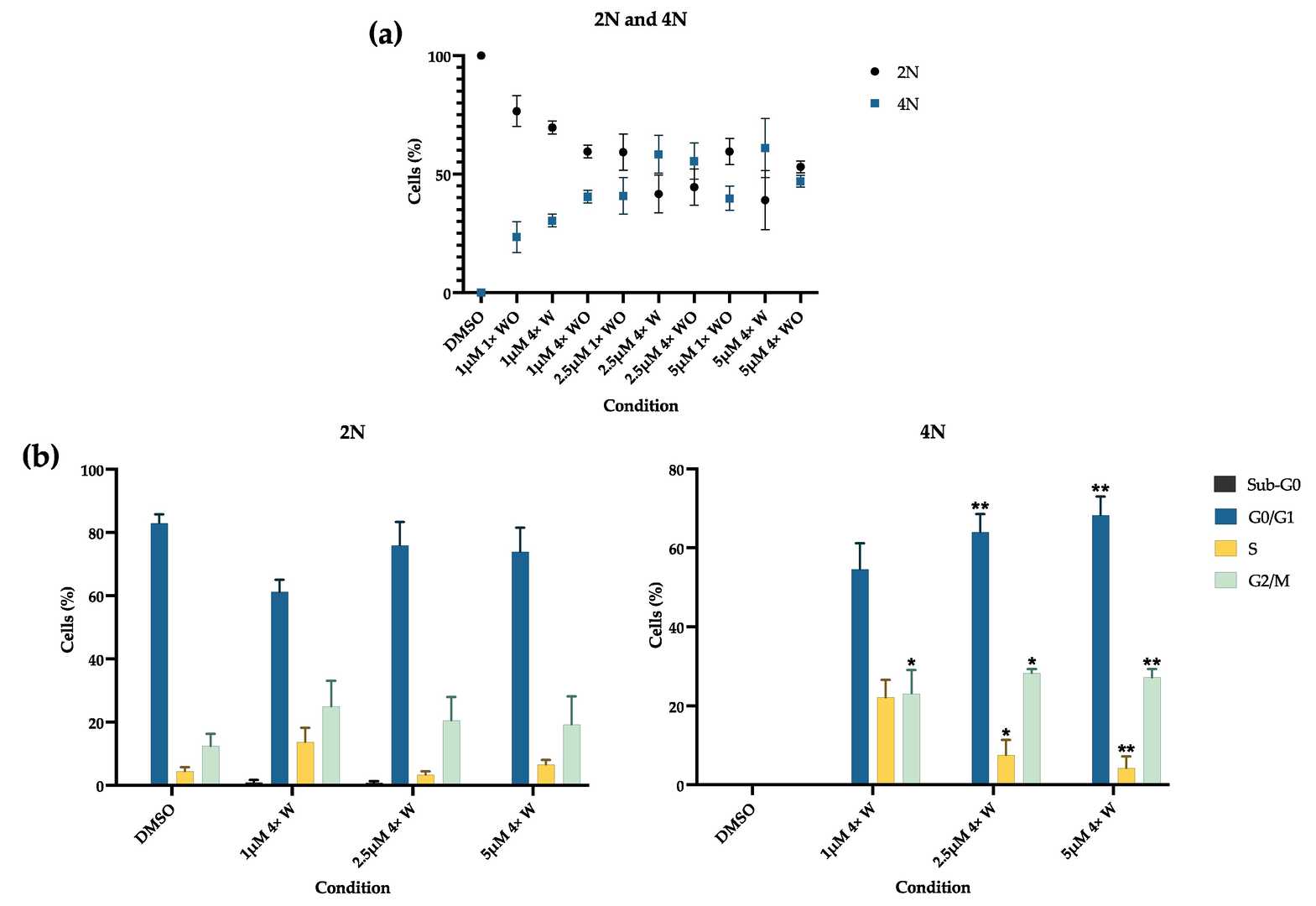 Fig. 1. Cell cycle analysis (Marto CM, Laranjo M, et al., 2024).
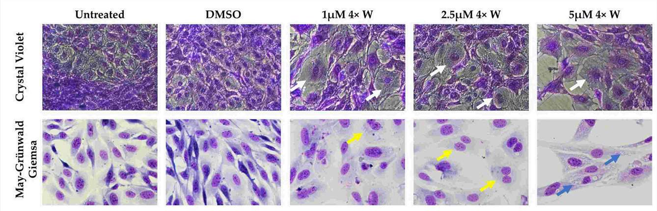 Fig. 2. Cell morphology before and after treatment (Marto CM, Laranjo M, et al., 2024).
Salicin Induces Taste Signaling via Tas2r143 in Mouse Gingival Fibroblasts (MGFs)
Periodontitis results in periodontal destruction due to dysbiotic biofilm buildup and immune response. Gingival fibroblasts, major components of gingiva, participate in immune responses through cytokine secretion. Recent studies suggest fibroblasts' taste receptor-like SCC activity helps regulate inflammation.
Zhang's team explored the potential of salicin to activate Tas2r143-mediated signaling in gingival fibroblasts to alleviate periodontitis. They investigated whether salicin was able to elicit calcium response via Tas2r143. They selected 10mM concentration of salicin with the most obvious calcium influx reaction for subsequent experiment by setting concentration gradients of 1,5,10 mM salicin. Single cell calcium imaging showed that salicin induced a strong increase of intracellular Ca2+ in MGFs (Fig. 3A). Consistently, an increase of intracellular Ca2+ was also detected in HEK293 cells with heterologous expression of Tas2r143 upon salicin stimulation (Fig. 3B). They further knocked down Tas2r143 in MGFs using siRNA, and the high efficiency of Tas2r143 gene silencing was observed through fluorescence microscopy and qRT-PCR. They found that the calcium flow response of MGFs to salicin was significantly weakened (Fig. 3C), indicating that salicin elicited taste-like response in MGFs via Tas2r143.
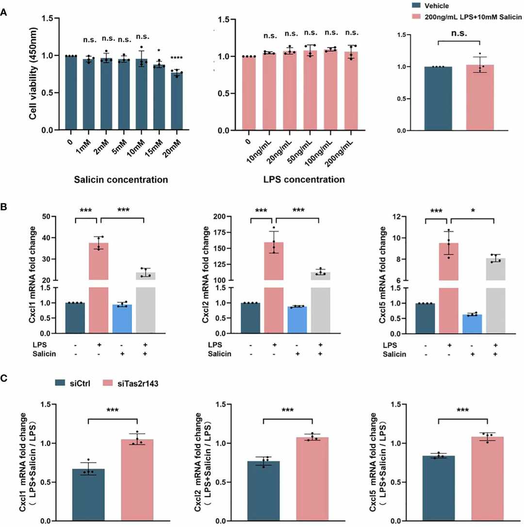 Fig. 3. Salicin inhibits LPS-induced chemokine expression by MGFs via Tas2r143 (Zhang Z, Zhou Z, et al.,2024).
Ask a Question
Write your own review
- You May Also Need

Description: C57BL/6-GFP Mouse Skeletal Muscle Microvascular Endothelial Cells from Creative Bioarray are isolated from C57BL/6-Tg (CAG-EGFP) 1Osb/J mouse skeletal muscle tissue of pathogen-free laboratory mice. ...

Description: eNOS KO Mouse Stomach Epithelial Cells from Creative Bioarray are isolated from stomach tissue of pathogen-free laboratory mice. eNOS KO Mouse Stomach Epithelial Cells are grown in a T25 tissue ...
Description: eNOS KO Mouse Liver Fibroblasts from Creative Bioarray are isolated from liver tissue of pathogen-free laboratory mice. eNOS KO Mouse Liver Fibroblasts are grown in T75 tissue culture flasks ...
Description: C57BL/6-GFP Mouse Corneal Epithelial Cells from Creative Bioarray are isolated from C57BL/6-GFP-Tg(CAG-EGFP)1Osb/J mouse corneal tissue of pathogen-free laboratory mice. C57BL/6-GFP Mouse Corneal ...

Description: BALB/c Mouse Retinal Microvascular Endothelial Cells from Creative Bioarray are isolated from retinal tissue of pathogen-free laboratory mice. BALB/c Mouse Retinal Microvascular Endothelial Cells are ...