
Rabbit Red Blood Cells
Cat.No.: CSC-C3103
Species: Rabbit
Source: Blood
Cell Type: Red Blood Cell
- Specification
- Background
- Scientific Data
- Q & A
- Customer Review
Rabbit Red Blood Cells (RBCs) are a primary type of cell in the blood, originating from the bone marrow. Bone marrow is divided into two types: red and yellow. And red marrow is involved in the production of red blood cells. During the development of rabbits, the production of red blood cells peaks within three weeks after birth, during which the bone marrow is in a state of high proliferation to meet the body's demand for blood components. The production of red blood cells is regulated by a variety of chemical factors, including those that promote red blood cell production and those involved in hemoglobin synthesis. Morphologically, rabbit red blood cells are similar to human red blood cells, being biconcave disc-shaped and lacking a nucleus. They are mainly involved in the transport of oxygen and carbon dioxide. In addition to the transport of gases, it is known that RBCs can function as immune molecules, for example, as antigen-presenting cells in immune responses.
Rabbit red blood cells are used in a variety of research studies, with their most common use in the determination of hemolytic activity of proteins or drugs. Red blood cells are also used in immunological studies, where they are often used in immune responses and antibody production. In cell biology, rabbit red blood cells are often used to study cell surface receptors, cell signaling, and other cell membrane functions.
FHR5 Decreases Hemolysis of Rabbit RBCs
Atypical hemolytic uremic syndrome (aHUS) can be linked to mutations/deletions/hybrid genes in factor H-related (FHR) proteins. Aradottir et al. examined a child with aHUS. Serum FHR5 was evaluated by immunoblotting, ELISA and by induction of rabbit red blood cell hemolysis in the presence/absence of recombinant human rFHR5. Combining rabbit RBCs with patient serum induced hemolysis which was reduced when recombinant hFHR5 was added at physiological concentrations (Fig. 1a). Similar results were seen when using serum from the father who has the CFHR5 M514R variant, without antibodies to factor H. The father's serum was also combined with hFHR5 at higher concentrations, also showing reduced hemolysis (Fig. 1b). Using control sera, the amount of hemolysis was decreased when adding high concentrations of hFHR5 but did not change significantly when adding physiological concentrations (Fig. 1). Adding hFHR5 to the cells before adding the serum, or combined with serum gave similar results.
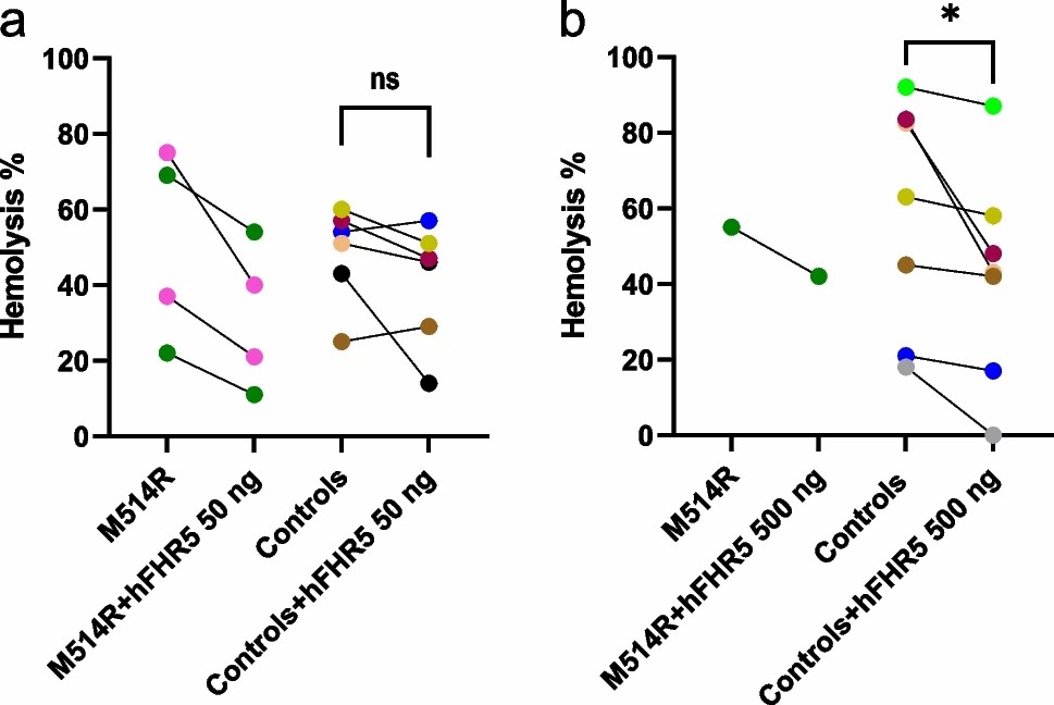 Fig. 1. Hemolysis induced by patient, father's and normal serum in the presence of factor H-related protein 5 (Aradottir S S., Kristoffersson AC, et al., 2024).
Evaluation of the Hemolytic Activity of rTβ4
The thymosin β4 from Pinctada fucata was expressed in this study, purified by Pichia pastoris expression system and Ni-NTA affinity chromatography. To assess the hemolytic activity of rTβ4, a 4% rabbit red blood cell suspension was used in an in vitro test. As seen in Figure 2, the positive control (1.0% Triton X-100) hemolysis rate was near to 100%, indicating that the experimental system was effective and sensitive. The negative control (PBS) showed a little hemolysis, indicating that the experimental system was stable. The hemolysis rate of rTβ4 protein was only 3.27% at 0.1 mg/mL, much lower than the safety threshold value of 5%. Meanwhile, hemolysis rate was unapparent at lower concentration (Fig. 2). Therefore, the results indicated that rTβ4 protein had good blood compatibility and could be used in further biomedical studies.
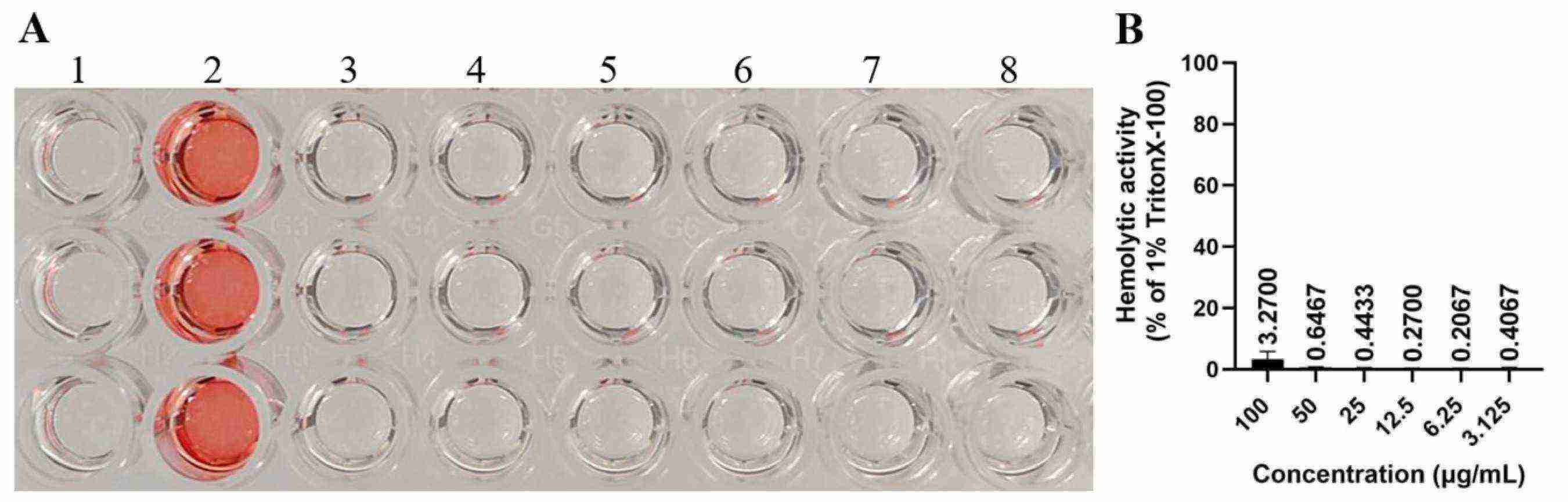 Fig. 2. In vitro hemolytic activity of rTβ4 protein (Liu P, Mo X, et al., 2025).
Ask a Question
Write your own review

Description: Rabbit Hepatocytes are derived from the liver of New Zealand White Rabbit.
Description: The aortic arch is the top part of the main artery carrying blood away from the heart. It is the connection between the ascending and descending aorta, and its central part is formed by the left 4th ...
Description: The synovium secretes synovial fluid, which plays an important role in joint movement. The normal synovium has two layers, a thin cellular layer (luminal layer) and a vascular layer (subintima). ...
Description: The pituitary gland is an important endocrine gland in the body that secretes growth hormone and adrenocorticotropic hormone. It plays an important role in the growth and development of the body, ...
Description: The oral epthelial cells are responsible for important functions, like the primary protection of oral mucosa against external aggressions building a mechanical barrier against microorganisms, ...
Description: The carotid arteries are major blood vessels in the neck that supply blood to the brain, neck, and face. There are two carotid arteries, one on the right and one on the left. In the neck, each ...