CCB
Cat.No.: CSC-C9042H
Species: Cyprinus carpio (Common carp)
Source: Brain
Morphology: Squamous
Culture Properties: Adherent
- Specification
- Background
- Scientific Data
- Q & A
- Customer Review
The CCB cell line is a fibroblast-like, diploid cell line originating from the brain of a Common carp (Cyprinus carpio carpio). It is a continuous and adherent cell line. CCB cells are noted for having healthy, robust growth in culture with typical elongated, fibroblastic morphology.
Biologically, the CCB cell line is most commonly used as a model for aquatic virology and fish health research. This line is highly susceptible to infection by many important viruses that infect fish, but most notably Cyprinid herpesvirus 3 (CyHV-3), Koi Herpesvirus (KHV). For this reason, CCB is the most commonly used cell line for in vitro study of viral replication cycle and pathogenesis, diagnostic test development and vaccine production against KHV, an economically important and often fatal disease. The cell line is also broadly used in toxicological applications to determine cytotoxicity and genotoxicity of various environmental pollutants such as heavy metals, pesticides and more to model the impact they have on aquatic species. This stable cell line is used in other comparative immunology studies as well to help to study and understand the mechanisms of innate immune responses of teleost fish. Gene expression studies, transfection experiments and other basic research of physiological mechanisms in cold-water fish species have also been done with CCB cells.
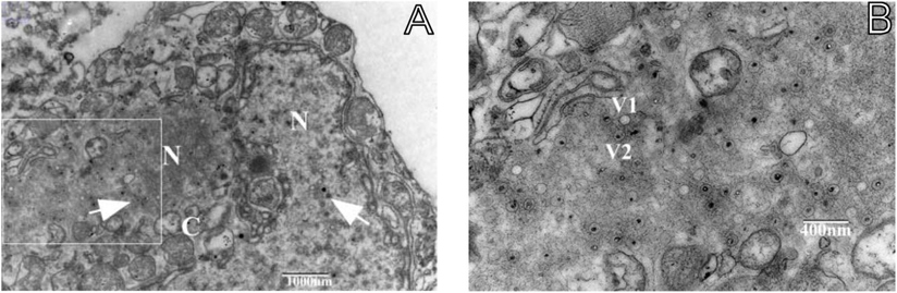
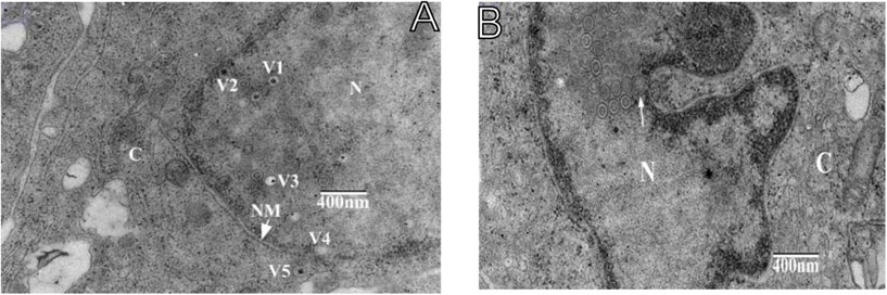
Electron Microscopic Observation of Infected CCB and Whole Genome Analysis of Koi Herpesvirus Isolate GY01
Cyprinid herpesvirus-3 (CyHV-3), also known as Koi herpesvirus (KHV), causes infectious and acute viremia in common and koi carp (Cyprinus carpio). In a previous study, a KHV isolate, GY1506 (KHV GY01 in GeneBank), was isolated from diseased common carp, cultured on CCB cells, and identified by PCR targeting the thymidine kinase (TK) gene and phylogenetic analysis. Further characterization and epidemiological features of this strain were studied through electron microscopy and whole-genome analysis.
Electron microscopy of CCB cells infected with KHV GY01 revealed incomplete nuclear membranes, deformed nuclei, marginalized nuclear chromosomes, and immature viruses arranged in a lattice pattern in the cytoplasm and nucleus (Fig. 1). Many mature and immature virus particles were observed. Three types of viruses were identified: hexagonal hollow structures, round capsules with dense internal substances, and clear visible structures of capsules, capsids, and cores. Viruses with or without capsules in GY01-infected CCB cells measured approximately 90nm by 110nm. A clear hexagonal virus was visible at the nuclear membrane edge (arrow). The cytoplasm protruded into a bubble (Fig. 2B).
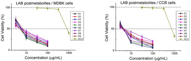
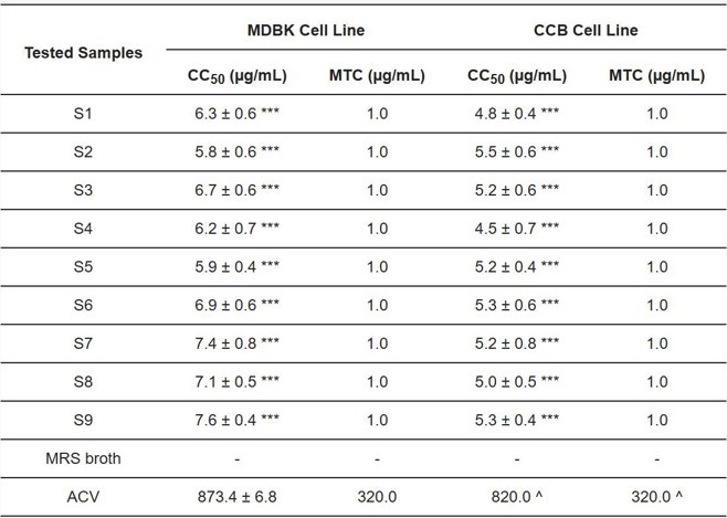
Lactobacilli-Derived Postmetabolites are Broad-Spectrum Inhibitors of Herpes Viruses In Vitro
Herpes viruses cause significant health, social, and economic losses across vertebrates. Lactic acid bacteria (LAB) are known to protect against pathogens through their metabolic compounds. Danova et al. evaluated the inhibitory properties of nine LAB postmetabolites against HSV-1 and KHV using cytotoxicity, cytopathic effect inhibition, virucidal effect, and protective effect assessments. They aim to explore the potential of LAB postmetabolites in preventing and treating herpes diseases.
Before the antiviral experiments, the non-toxic range of each sample was determined on Madin-Darby bovine kidney (MDBK) and common carp brain (CCB) cell lines. Cytotoxicity was measured at 48 hours on MDBK cells and at 72 hours on CCB cells, following the antiviral evaluation protocol (Fig. 3). Figure 4 shows that all postbiotics were more toxic than the reference chemotherapeutic acyclovir (ACV). Cytotoxicity was slightly higher in CCB cells, likely due to 24 hours longer exposure (72 hours) compared to MDBK cells (48 hours). The Mix-sample group (S7, S8, and S9) had the lowest toxicity. S9 was the least cytotoxic, followed by S6, S3, S1, and S4. S5 and S2 showed the highest cytotoxicity (Fig. 4).
Ask a Question
Write your own review
- You May Also Need


Description: Derived from tissue posterior to the anus of normal adult minnows. Reported to be capable of growth over a wide temperature range from 4°C-36°C with maximum growth at 34°C. Support growth of the ...
Description: E11 is a clone of the cell line SSN-1 and is persistantly infected with a C-type retrovirus (SnRV). It is susceptible to piscine nodavirus strains belonging to different genotypes (SJNNV, RGNNV, ...

Description: The fish cell line SSN-1 was initiated from whole fry tissue, Channa (Ophicephalus) striatus, commonly named 'striped snakehead'. SSN-1 spontaneously produce and release endogenous snakehead fish ...

Description: From pooled male/female gonad tissue of yearling rainbow trout (Oncorhynchus mykiss). First cell line to be established from cold-blooded vertebrates. 8-fold difference in growth rate in temperature ...

- Adipose Tissue-Derived Stem Cells
- Human Neurons
- Mouse Probe
- Whole Chromosome Painting Probes
- Hepatic Cells
- Renal Cells
- In Vitro ADME Kits
- Tissue Microarray
- Tissue Blocks
- Tissue Sections
- FFPE Cell Pellet
- Probe
- Centromere Probes
- Telomere Probes
- Satellite Enumeration Probes
- Subtelomere Specific Probes
- Bacterial Probes
- ISH/FISH Probes
- Exosome Isolation Kit
- Human Adult Stem Cells
- Mouse Stem Cells
- iPSCs
- Mouse Embryonic Stem Cells
- iPSC Differentiation Kits
- Mesenchymal Stem Cells
- Immortalized Human Cells
- Immortalized Murine Cells
- Cell Immortalization Kit
- Adipose Cells
- Cardiac Cells
- Dermal Cells
- Epidermal Cells
- Peripheral Blood Mononuclear Cells
- Umbilical Cord Cells
- Monkey Primary Cells
- Mouse Primary Cells
- Breast Tumor Cells
- Colorectal Tumor Cells
- Esophageal Tumor Cells
- Lung Tumor Cells
- Leukemia/Lymphoma/Myeloma Cells
- Ovarian Tumor Cells
- Pancreatic Tumor Cells
- Mouse Tumor Cells