
C57BL/6 Mouse Primary Mammary Epithelial Cells
Cat.No.: CSC-C4262X
Species: Mouse
Source: Breast
Cell Type: Epithelial Cell
- Specification
- Background
- Scientific Data
- Q & A
- Customer Review
Mouse Primary Mammary Epithelial Cells can be used in assays of cell to cell adhesion and migration. Standard biochemical procedures performed with epithelial cell cultures include RT-PCR, Western blotting, immunoprecipitation, immunofluorescent staining or immunofluorescent flow cytometry or generating cell derivatives for desired research applications.
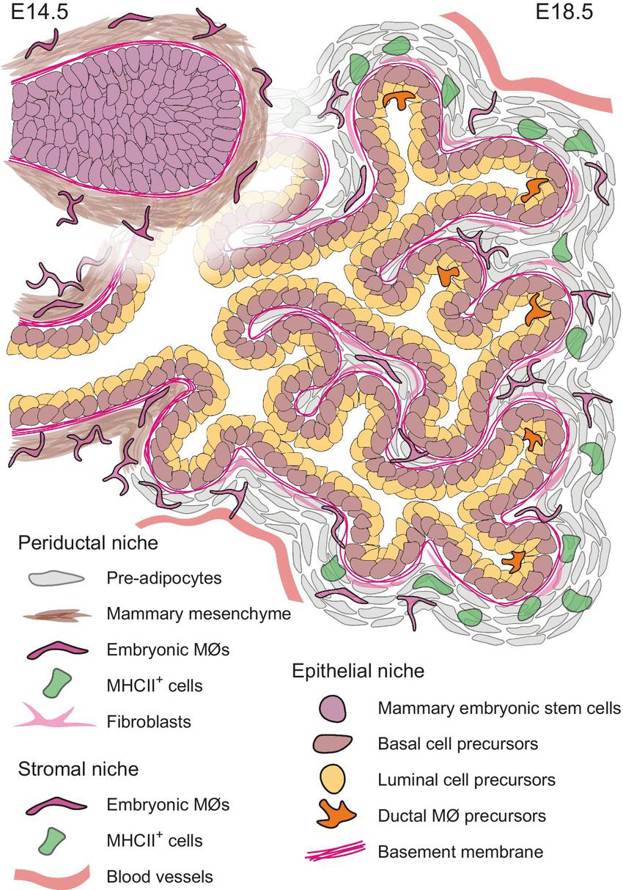
Creative Bioarray's mouse primary mammary epithelial cells are sourced from the mammary tissue of healthy C57BL/6 mice. C57BL/6 mice are a popular inbred strain that have been well characterized, making them a common and reliable choice for laboratory mice. The mammary gland is a complex organ composed of epithelial cells, which are responsible for milk production during lactation. Mammary epithelial cells are found in the ducts and alveoli of the mammary gland. Morphologically, mammary epithelial cells have the typical appearance of epithelial cells, including a cobblestone-like appearance in monolayer cultures. They also express epithelial cell-specific markers, such as E-cadherin and ZO-1, which can be detected using immunofluorescent staining.
Mammary epithelial cells play an important role in the development and function of the mammary gland. During puberty, they are involved in the formation of the ductal tree. During pregnancy, mammary epithelial cells differentiate into alveolar cells and produce milk during lactation. These cells are ideal for investigating mammary gland development, differentiation, and pathology, providing a reliable model for both basic and translational research.
L-arginine Promotes Cell Proliferation by Up-Regulating the Expression of Proliferation-Related Proteins
Amino acids have been shown to affect the development of mammary gland (MG). However, it is unclear whether L-arginine promotes the development of pubertal MG. Therefore, Ge's team aims to explore the effect of L-arginine on the development of MG in pubertal mice.
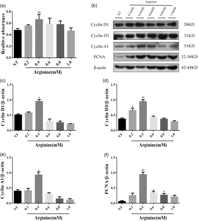
To investigate its internal mechanism of action, they used mouse mammary epithelial cells (mMECs) line. The results revealed that L-arginine can significantly promote the development of MG. Next, they investigated the effect of various concentrations of L-arginine (0, 0.2, 0.4, 0.6, 0.8 and 1.0 mM) on the proliferation of mMECs. The results showed that mMECs had the most obvious effect on their proliferation after being treated with 0.4 mM L-arginine (Fig. 1a). Furthermore, L-arginine significantly increased the protein levels of proliferation-related proteins such as Cyclin D1/D3/A1 and PCNA in mMECs (Fig. 1b-f). The final results indicate that L-arginine promotes the proliferation of mMECs and increases the levels of proliferation-related proteins.
BMP4 Differentiates Mammary Epithelial Cells into an Acinar Structure in 3D Cell Culture
Basal-like triple-negative breast cancer (TNBC) is characterized by an aggressive phenotype with poor prognosis, relapse, and drug resistance mediated by a high cancer stem-cell (CSC) burden. The mechanisms sustaining the stemness feature in this breast cancer subtype are not well understood. Here, Yan et al. asked how TGFβ family ligands network to control CSC abundance in TNBC, and whether opposing factors could be therapeutically exploited.
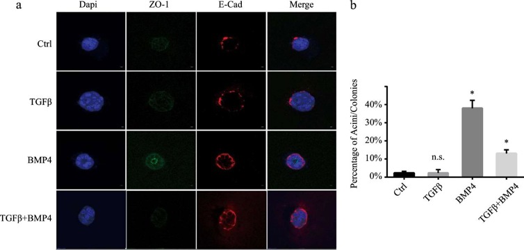
They evaluated whether BMP4 could induce the formation of mammary acinar structures in normal mouse mammary epithelial cells. They performed ex vivo acini morphogenesis assays in primary mammary epithelial cells isolated from female virgin mice. As seen in Fig. 2a, BMP4 strongly induced the formation of organized mammary acini with well-established apical/basal polarity (as indicated by the apical localization of ZO-1 and basal/lateral localization of E-cadherin) compared to the control and TGFβ-stimulated cells that did not form such organized acini. Interestingly, TGFβ strongly antagonized the effects of BMP4 on acinar morphogenesis. They then quantified the number of acini under different conditions. As shown in Fig. 2b, BMP4 significantly increased the efficiency of acinar formation, an effect that was antagonized by TGFβ. These data point to BMP4 as a strong differentiation factor in normal mammary epithelial cells that efficiently promotes the formation of well-organized 3D acinar structures, and TGFβ can efficiently antagonize this BMP4 activity.
Ask a Question
Write your own review
Description: C57BL/6-GFP Mouse Skeletal Muscle Microvascular Endothelial Cells from Creative Bioarray are isolated from C57BL/6-Tg (CAG-EGFP) 1Osb/J mouse skeletal muscle tissue of pathogen-free laboratory mice. ...

Description: eNOS KO Mouse Stomach Epithelial Cells from Creative Bioarray are isolated from stomach tissue of pathogen-free laboratory mice. eNOS KO Mouse Stomach Epithelial Cells are grown in a T25 tissue ...
Description: eNOS KO Mouse Liver Fibroblasts from Creative Bioarray are isolated from liver tissue of pathogen-free laboratory mice. eNOS KO Mouse Liver Fibroblasts are grown in T75 tissue culture flasks ...
Description: C57BL/6-GFP Mouse Corneal Epithelial Cells from Creative Bioarray are isolated from C57BL/6-GFP-Tg(CAG-EGFP)1Osb/J mouse corneal tissue of pathogen-free laboratory mice. C57BL/6-GFP Mouse Corneal ...

Description: BALB/c Mouse Retinal Microvascular Endothelial Cells from Creative Bioarray are isolated from retinal tissue of pathogen-free laboratory mice. BALB/c Mouse Retinal Microvascular Endothelial Cells are ...