Immortalized Human Skin Fibroblast-hTERT
Cat.No.: CSC-I9069L
Species: homo sapiens
Source: Skin
Morphology: Bipolar
Culture Properties: Adherent
- Specification
- Background
- Scientific Data
- Q & A
- Customer Review
Note: Never can cells be kept at -20 °C.
CIK-HT013 HT® Lenti-hTERT Immortalization Kit
free from contaminations (bacteria incl. mycoplasma, fungi, HIV, HAV, HBV, HCV, Parvo-B19) and cross-contaminations
Skin fibroblasts are essential to skin biology, playing key roles in extracellular matrix production, wound healing, and skin regeneration. Primary fibroblasts have long been the gold standard model system for studying skin biology, however, donor variability and the limited lifespan of primary cells have limited their potential.
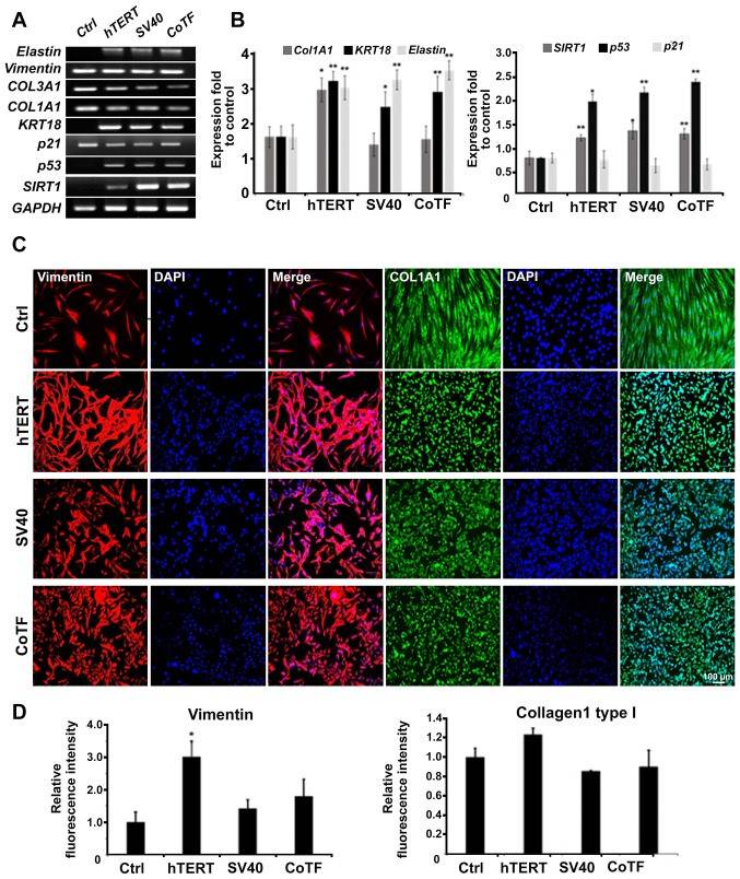
While traditional methods of immortalizing primary cells using SV40 can overcome these limitations, they often result in loss of normal biological functions. hTERT can fully immortalize human skin fibroblasts while retaining the physiological properties of primary cells, providing an unlimited source of cells for research on skin diseases, skin aging, wound healing, cosmetics, and toxicology, and helping to improve research reproducibility.

EBOV Infection of Primary and Immortalized Human Skin Fibroblasts
Ebola virus (EBOV) causes severe human disease. During late infection, EBOV virions are on the skin s surface, however, the permissive skin cell types and the route of virus translocation to the epidermal surface are unknown.
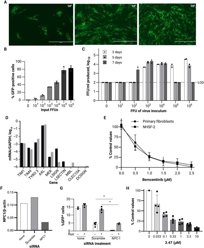
This study evaluated EBOV infection in cultured primary and immortalized human skin fibroblasts (NHSF-2) and identified host receptors that mediate virus entry. Primary skin human fibroblasts were infected with EBOV and a dose-dependent increase in virus infection was evident on day 5 (Fig. 1A) and day 7 (Fig. 1B). Gene expression studies in the NHSF-2 cells demonstrated that AXL was more than 100-fold more highly expressed than other known cell surface receptors used by EBOV (Fig. 1D) and treatment of NHSF-2 cells or a primary skin fibroblast population with the AXL inhibitor, bemcentinib, inhibited virus infection (Fig. 1E), indicating that AXL is critical for EBOV entry into fibroblasts. Last, the requirement for the endosomal receptor NPC1 was evaluated in siRNA knockdown and 3.47 inhibition studies. Ruxolitinib enhanced virus infection in the presence of the scrambled siRNA but, as anticipated, decreased rVSV/EBOV GP infection upon NPC1-specific siRNA transfection (Fig. 1, F and G). Treatment of NHSF-2 with NPC1 inhibitor 3.47 also resulted in decreased EBOV GP dependent entry (Fig. 1H). In total, these studies demonstrate that skin fibroblasts support EBOV infection in an AXL-dependent and NPC1-dependent manner.

Ask a Question
Write your own review
- Adipose Tissue-Derived Stem Cells
- Human Neurons
- Mouse Probe
- Whole Chromosome Painting Probes
- Hepatic Cells
- Renal Cells
- In Vitro ADME Kits
- Tissue Microarray
- Tissue Blocks
- Tissue Sections
- FFPE Cell Pellet
- Probe
- Centromere Probes
- Telomere Probes
- Satellite Enumeration Probes
- Subtelomere Specific Probes
- Bacterial Probes
- ISH/FISH Probes
- Exosome Isolation Kit
- Human Adult Stem Cells
- Mouse Stem Cells
- iPSCs
- Mouse Embryonic Stem Cells
- iPSC Differentiation Kits
- Mesenchymal Stem Cells
- Immortalized Human Cells
- Immortalized Murine Cells
- Cell Immortalization Kit
- Adipose Cells
- Cardiac Cells
- Dermal Cells
- Epidermal Cells
- Peripheral Blood Mononuclear Cells
- Umbilical Cord Cells
- Monkey Primary Cells
- Mouse Primary Cells
- Breast Tumor Cells
- Colorectal Tumor Cells
- Esophageal Tumor Cells
- Lung Tumor Cells
- Leukemia/Lymphoma/Myeloma Cells
- Ovarian Tumor Cells
- Pancreatic Tumor Cells
- Mouse Tumor Cells
