
Immortalized Mouse Epidermal Cells (COCA)
Cat.No.: CSC-I9267L
Species: Mus musculus
Source: Skin
Morphology: Polygonal
Culture Properties: Adherent
- Specification
- Background
- Scientific Data
- Q & A
- Customer Review
Note: Never can cells be kept at -20 °C.
2) ELISA was used to determine the TNFα secretion after inflammatory stimulation;
3) Western blot and immunocytochemistry were used to confirm the expression of microglial specific markers such as CD68 and Iba1 and M1/M2 phenotype-associated protein expression (COX-2/iNOS and Arg-1) after stimulation;
4) Aβ1-42orE. coli-derived bioparticles uptake was used to determine phagocytic activity of SIM-A9 cells.
Immortalized Mouse Epidermal Cells (COCA) is a spontaneously immortalized murine keratinocyte cell line derived from primary epidermal keratinocytes collected from the dorsal skin of adult wildtype (C57BL/DBA) mice. COCA was derived by continuously culturing primary mouse keratinocytes in defined, low‑calcium medium lacking feeder layers or serum allowing for stable long‑term propagation (>75 passages) without apparent loss of physiologic epidermal properties. Morphologically epithelial, COCA cells undergo calcium‑induced differentiation and can form stratified epidermis in vitro (2D and 3D). COCA express keratinocyte‑specific markers such as K5, K10, loricrin, and filaggrin and can reestablish normal epidermal differentiation programs. Cells are non‑tumorigenic and able to regenerate morphologically normal epidermis when grafted in vivo. For these reasons COCA serves as an excellent physiologically relevant in vitro model system to study epidermal biology, skin barrier formation, wound repair mechanisms, three-dimensional skin tissue engineering, as well as pathogenesis of skin diseases.
Serum Affects Keratinization and Tight Junctions in 3D Cultures of the Mouse Keratinocyte Cell Line COCA through Retinoic Acid Receptor-Mediated Signaling
Vitamin A in serum affects keratinocyte proliferation, differentiation, and keratinization. While mouse oral, esophageal, and forestomach epithelia are keratinized, human equivalents are not. Ozaki et al. evaluated serum effects on morphology, differentiation markers, tight junction proteins, and paracellular permeability in 3D cultures of mouse keratinocytes.
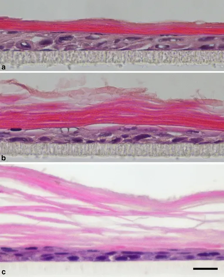
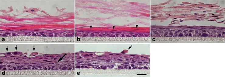
A new 3D medium (CnT-PR 3D Barrier) replaced the previous formulation, which did not induce keratinized stratified epithelium-like structures in COCA cultures. Epidermal analogs were generated by adding calcium, KGF, and APM to CnT-PR. COCA formed epidermis-like structures within 1 week, maintained for at least 3 weeks, though the cornified layer became loosely packed (Fig. 1). Serum effects were examined after 2 weeks (Fig. 2), given its essential role in skin equivalent models. Control cultures formed keratinized stratified squamous epithelium (SSE) (Fig. 2a). With 0.01% charcoal-stripped FBS (ch-FBS), nuclei persisted in the cornified layer (Fig. 2b, arrowheads). Keratinization was inhibited by 0.1%, 1%, and 10% ch-FBS (Fig. 2c-e). Non-keratinized SSE with 0.1% ch-FBS showed 2-3 superficial squamous cell layers with detached nucleated cells (Fig. 2c). Hyperplastic rather than flat superficial cells appeared with 1% or 10% ch-FBS, with some detaching (Fig. 2d, e, arrows). Flattened cell layers were observed beneath hyperplastic surfaces (Fig. 2d, large arrow), and large intercellular gaps occurred at 10% ch-FBS (Fig. 2e).
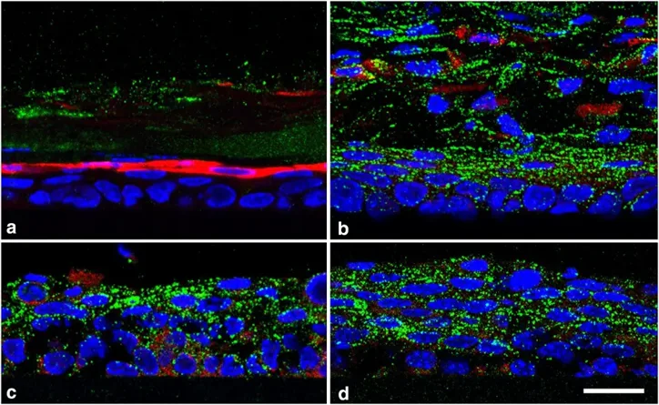
Immunofluorescence revealed LOR (late keratinization marker) in cells beneath the cornified layer without ch-FBS, but K4 (non-keratinization marker) was absent from nucleated cells (Fig. 3a). With ≥0.1% ch-FBS, K4 replaced LOR in all suprabasal cells of non-keratinized SSE (Fig. 3b-d). Antibody specificity was confirmed in porcine oral mucosa.
Ask a Question
Write your own review
Description: Immortalized Mouse Primary Artery Fibroblasts-GFP provided by Creative Bioarray have been developed by immortalizing mouse artery fibroblasts with SV40 Large T antigen and transfecting with tGFP. The ...

Description: Immortalized MHC II -/- Mouse Macrophage Cells (C2D) is derived from the knockout mice negative for MHCII molecule. They are stable in culture and survived through crisis in which they spontaneously ...
Description: The bone morphogenetic proteins (BMPs) produced by calvarial cells are crucial in osteoblastic differentiation and bone regeneration, however the limited lifespan of primary cells diminishes their ...

Description: The Immortalized Mouse Atrioventricular Cushion Mesenchymal Cells (tsA58-AVM) were derived from atrioventricular (AV) cushions of H-2Kb -tsA58 embryos at E9.5 and were conditionally immortalized in ...
Description: IDG-SW3 represent a non-homogenous population progressing from early osteoblasts to late osteocytic. These cells express functional SV40 large T antigen that is induced in the presence of IFN γ under ...
Description: The Immortalized Mouse Spleen Dendritic Cell (SRDC) line is functionally and phenotypically similar to dendritic cells; specifically, in its antigen presentation, T cell priming, and dendritic cell ...
- Adipose Tissue-Derived Stem Cells
- Human Neurons
- Mouse Probe
- Whole Chromosome Painting Probes
- Hepatic Cells
- Renal Cells
- In Vitro ADME Kits
- Tissue Microarray
- Tissue Blocks
- Tissue Sections
- FFPE Cell Pellet
- Probe
- Centromere Probes
- Telomere Probes
- Satellite Enumeration Probes
- Subtelomere Specific Probes
- Bacterial Probes
- ISH/FISH Probes
- Exosome Isolation Kit
- Human Adult Stem Cells
- Mouse Stem Cells
- iPSCs
- Mouse Embryonic Stem Cells
- iPSC Differentiation Kits
- Mesenchymal Stem Cells
- Immortalized Human Cells
- Immortalized Murine Cells
- Cell Immortalization Kit
- Adipose Cells
- Cardiac Cells
- Dermal Cells
- Epidermal Cells
- Peripheral Blood Mononuclear Cells
- Umbilical Cord Cells
- Monkey Primary Cells
- Mouse Primary Cells
- Breast Tumor Cells
- Colorectal Tumor Cells
- Esophageal Tumor Cells
- Lung Tumor Cells
- Leukemia/Lymphoma/Myeloma Cells
- Ovarian Tumor Cells
- Pancreatic Tumor Cells
- Mouse Tumor Cells