MM.1S
Cat.No.: CSC-C9219W
Species: Homo sapiens (Human)
Source: Blood; Peripheral Blood
Morphology: lymphoblast
- Specification
- Background
- Scientific Data
- Q & A
- Customer Review
- Documents
The MM.1S cell line is a widely used and well-characterized cell line derived from a patient with multiple myeloma (MM), a cancer of plasma cells in the bone marrow. It has since become a valuable tool for studying the biology of MM and evaluating potential therapeutic strategies. The MM.1S cell line exhibits several characteristic features of MM, including the ability to produce monoclonal immunoglobulin and express plasma cell markers such as CD138.
The MM.1S cell line has been extensively characterized and extensively used in research related to MM. It has contributed to our understanding of the molecular mechanisms underlying the development and progression of MM, as well as the evaluation of novel therapeutic agents and treatment approaches. Researchers often utilize the MM.1S cell line to investigate various aspects of MM, including cell signaling pathways, drug sensitivity, genetic alterations, and interactions with the bone marrow microenvironment.
Characterization of the MM.1 Cell Lines
MM is a clonal B-lymphocyte malignancy characterized by accumulating terminally differentiated antibody-producing cells in the bone marrow. The MM.1 cells grow in suspension or lightly attached to the flask singly or in small clusters. Their growth media is supplemented only by 10% fetal calf serum, glutamine, fungizone, and penicillin-streptomycin. The cells have a doubling time of 72 hours and grow best at high density.
The MM.1 cells have typical myeloma cell morphology. They are round with eccentrically located nuclei, many of which are binucleated or multinucleated (Fig. 1). They have well-developed rough endoplasmic reticulum and prominent nucleoli. Cell staining showed that MM.1 was a cytochemical characteristic of myeloma cells. Immunocytochemical staining showed that the MM.1 cells contain cytoplasmic λ- and not a κ-light chain (Table 1).
The MM.1 cells are characteristic plasma cells. They are negative for CD19 and CD20 found on most normal and malignant B lymphocytes. They have low expression of CD10 (common lymphoblastic leukemia antigen), which is found on immature B cells, and have low expression of CD45. The MM.1 cells express the plasma cell surface marker CD38 (Table 1) and the plasma cell–associated antigen (PCA-1). The MM.1 cells also express HLA-DR (D-related human leukocyte antigen); CD59, which is a GPI-linked complement receptor seen on the surface of some lymphocytes; CD52; and CD25, the interleukin-2 (IL-2) receptor.
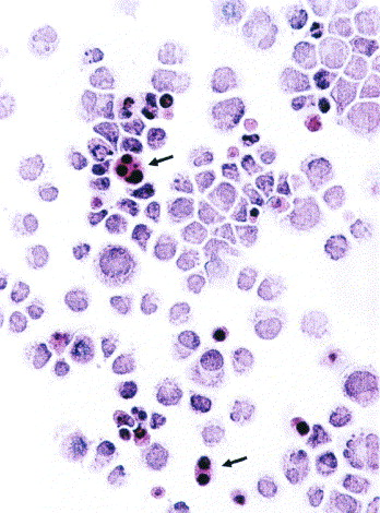 Fig. 1 Morphological features of the MM.1S cultured cells. (Greenstein S, et al., 2003)
Table 1. Expression of cell surface markers in the MM.1S cell lines (Greenstein S. et al., 2003).
| Cell surface marker | λ light chain | κ light chain | CD38 | CD10 | CD19 | CD20 | CD25 | CD45 | CD52 | CD59 |
| MM1.S | 92.3% | 0.2% | 96.1% | 0.3% | 0.2% | 0.2% | 79.0% | 0.3% | 98.1% | 95.7% |
TGF-β Induces Growth Suppression in MM.1S Cells
Transforming growth factor-β (TGF-β) is important in essential cell phenotypes, particularly in cell proliferation, differentiation, and apoptosis. The present study found that cell proliferation was markedly inhibited in TGF-β-treated MM.1S cells.
MM.1S cells were grown in the presence or absence of TGF-β. Cell viability was assessed using an MTS assay. It was found that the growth of MM.1S cells was inhibited by TGF-β in a time-dependent manner (P<0.05; Fig. 2A). This was confirmed by a reduction in cell count (P<0.05; Fig. 2B). MM.1S cells were seeded overnight at 3×10⁵/ml, during which time almost 60% of cells were synchronized in the G1 phase of the cell cycle, as measured by PI staining (Fig. 2C). Cells were then stimulated or not with TGF-β for 48 h. Control cells progressed from G1 to S phase through the cell cycle, whereas TGF-β-treated cells exhibited efficient G1 cell cycle suppression (P<0.05; Fig. 2C). The present study found that cell proliferation was markedly inhibited in TGF-β-treated MM.1S cells.
To study the contribution of E2F1 in mediating this TGF-β response, RNA interference was used to reduce the expression of endogenous E2F1. It was found that the effect of TGF-β on cell viability in the MM.1S cell lines tested was almost completely prevented when E2F1 expression was silenced, indicating that E2F1 is required for mediating the TGF-β-induced response in the MM.1S cell line (P<0.05; Fig. 3).
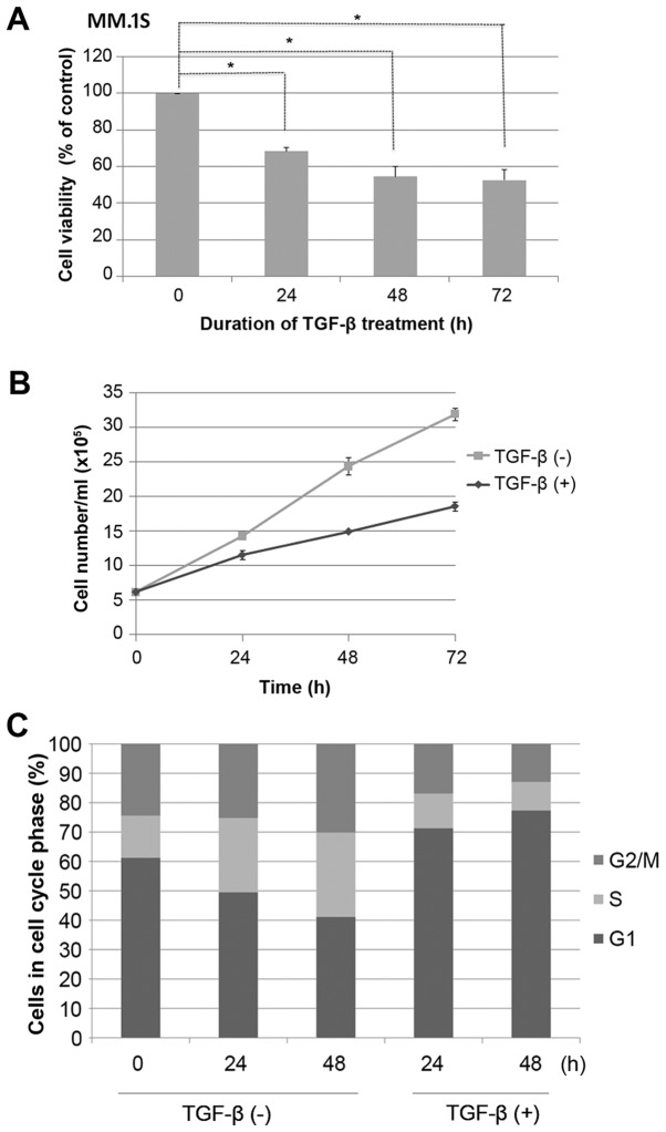 Fig. 2 TGF-β induces efficient G1 cell cycle suppression in MM.1S cells. (Liu X, et al., 2017)
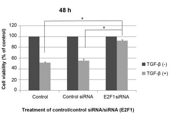 Fig. 3 TGF-β-mediated cell growth suppression is impaired in E2F1-null MM.1S cells. (Liu X, et al., 2017)
Ask a Question
Write your own review
- You May Also Need

Description: Established in 2007 from the large retrosternal mass resected before treatment from a 57-year-old Caucasian man with rapidly fatal anaplastic thyroid cancer

Description: established from the uvea tissue of a male patient with uveal melanoma

Description: NTERA-2 was cloned from cell line TERA-2 which was derived from a metastatic teratocarcinoma of a 22-year-old Caucasian male; cell line also known as NT-2

Description: Established from the bone marrow of a 27-year-old woman with B-cell non-Hodgkin lymphoma (B-NHL) (diffuse large cell lymphoma, DLCL, stage 4B, at relapse) in 1987
- Adipose Tissue-Derived Stem Cells
- Human Neurons
- Mouse Probe
- Whole Chromosome Painting Probes
- Hepatic Cells
- Renal Cells
- In Vitro ADME Kits
- Tissue Microarray
- Tissue Blocks
- Tissue Sections
- FFPE Cell Pellet
- Probe
- Centromere Probes
- Telomere Probes
- Satellite Enumeration Probes
- Subtelomere Specific Probes
- Bacterial Probes
- ISH/FISH Probes
- Exosome Isolation Kit
- Human Adult Stem Cells
- Mouse Stem Cells
- iPSCs
- Mouse Embryonic Stem Cells
- iPSC Differentiation Kits
- Mesenchymal Stem Cells
- Immortalized Human Cells
- Immortalized Murine Cells
- Cell Immortalization Kit
- Adipose Cells
- Cardiac Cells
- Dermal Cells
- Epidermal Cells
- Peripheral Blood Mononuclear Cells
- Umbilical Cord Cells
- Monkey Primary Cells
- Mouse Primary Cells
- Breast Tumor Cells
- Colorectal Tumor Cells
- Esophageal Tumor Cells
- Lung Tumor Cells
- Leukemia/Lymphoma/Myeloma Cells
- Ovarian Tumor Cells
- Pancreatic Tumor Cells
- Mouse Tumor Cells