
CT26.WT[CT26WT]
Cat.No.: CSC-C9171W
Species: Mus musculus (Mouse)
Source: Intestine; Colon
Morphology: fibroblast
- Specification
- Background
- Scientific Data
- Q & A
- Customer Review
CT26WT is a mouse colon carcinoma cell line. The parental line was generated by N‑nitroso‑N‑methylurethane (NNMU) chemical induction of primary undifferentiated colon tumors in BALB/c mice followed by isolation of a wild‑type clone. Cells are adherent with fibroblast‑like morphology, characterized by spindle‑shaped or polygonal bodies with relatively uniform nuclei. Under normal culture conditions (RPMI‑1640 + 10 % fetal bovine serum, 37 °C, 5 % CO₂), CT26WT has a rapid growth rate with doubling time of about 12-18 hours, reaching confluency of 70-80 %.
CT26WT is syngeneic in BALB/c mice and highly tumorigenic. A subcutaneous injection of 1 × 10⁶ cells results in palpable tumors within 8 days and of ~2 cm³ in size within about 2 weeks. Intravenous inoculation results in heavy lung colonization, and this property has been used as a model of metastatic spread. The parental line, as well as derivative clones (most prominently CT26.CL25, a stable clone expressing β‑galactosidase as a model tumor antigen) have been broadly used in immuno‑oncology research, including vaccine design, checkpoint‑inhibitor screening, and adoptive cell‑therapy studies.
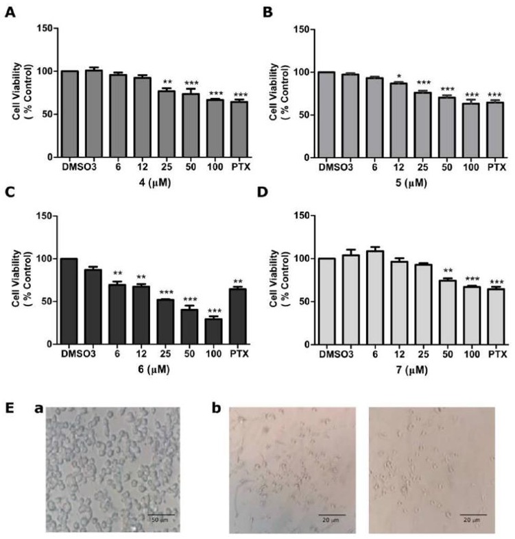
Antiproliferative Activity of Compounds 4, 5, 6, and 7 on CT26WT Cells
Colorectal cancer (CRC) is one of the most-deadly malignancies globally. Although, when resectable, surgery may offer patients prolonged survival, chemotherapy plays an important role in the management of metastatic CRC. Anticancer drugs currently in clinical use lack selectivity and are affected by cancerous mutations, therefore new chemotherapeutic molecules are urgently needed. Pacheco et al. report the synthesis, characterization, and antiproliferative activity of a series of four steroidal carbamates, in mouse colon carcinoma CT26WT cells.
The cytotoxicity of compounds 4, 5, 6 and 7, was determined in CT26WT cells by visualizing alterations in the cell morphology and by MTT reduction, to quantitate the antiproliferative effect after 24 h of incubation with the drugs. As can be seen in Fig. 1, all steroids inhibited the growth of CT26WT cells in a dose-dependent manner and were statistically different from untreated cells. The initial inhibitory concentrations of compounds 4, 5, 6 and 7 were found to be 25 μM, 12 μM, 6 μM and 50 μM, respectively (Fig. 1A-D). At a concentration of 25 μM, the inhibitory activity order was: 7 < 4 = 5 < 6. The most active compound (6) presented an IC50 of 26.8 μM, which is in accordance with the literature-reported values for steroidal carbamates. Morphologically, the number of shrunken, rounded and detached cells was higher when incubated with increasing concentrations of 4-7 compounds and not present in control cells (Fig. 1Ea, b).
Ask a Question
Write your own review
- You May Also Need

Description: Described as secreting a mouse monoclonal antibody (IgG2a) detecting all fibers in skeletal muscle and myosin heavy chains on Western blots and detecting mammalian, chicken, zebrafish, axolotl, ...

Description: Animals were immunized with the B6.1 mouse cytotoxic T cell line.


Description: neuroglial and neuronal character coexpressing ependymoma cell line.

Description: Established by irradiation of the adherent cells in long-term bone marrow cultures derived from C3H/HeNSlc strain mice
- Adipose Tissue-Derived Stem Cells
- Human Neurons
- Mouse Probe
- Whole Chromosome Painting Probes
- Hepatic Cells
- Renal Cells
- In Vitro ADME Kits
- Tissue Microarray
- Tissue Blocks
- Tissue Sections
- FFPE Cell Pellet
- Probe
- Centromere Probes
- Telomere Probes
- Satellite Enumeration Probes
- Subtelomere Specific Probes
- Bacterial Probes
- ISH/FISH Probes
- Exosome Isolation Kit
- Human Adult Stem Cells
- Mouse Stem Cells
- iPSCs
- Mouse Embryonic Stem Cells
- iPSC Differentiation Kits
- Mesenchymal Stem Cells
- Immortalized Human Cells
- Immortalized Murine Cells
- Cell Immortalization Kit
- Adipose Cells
- Cardiac Cells
- Dermal Cells
- Epidermal Cells
- Peripheral Blood Mononuclear Cells
- Umbilical Cord Cells
- Monkey Primary Cells
- Mouse Primary Cells
- Breast Tumor Cells
- Colorectal Tumor Cells
- Esophageal Tumor Cells
- Lung Tumor Cells
- Leukemia/Lymphoma/Myeloma Cells
- Ovarian Tumor Cells
- Pancreatic Tumor Cells
- Mouse Tumor Cells