CCD 841 CON
Cat.No.: CSC-C9165W
Species: Homo sapiens (Human)
Source: Intestine; Colon
Morphology: epithelial
- Specification
- Background
- Scientific Data
- Q & A
- Customer Review
The CCD 841 CoN cell line was derived from normal human colonic epithelial tissue from the colon of a 21-week-old female fetus. These cells are epithelial-like, adherent, and demonstrate disorganized, flattened growth with distinct clusters of round cells on the surface. However, the phenotype of this cell line has been called into question, and may not be representative of epithelial cells. Phenotypically, this cell line is negative for the epithelial differentiation marker cytokeratin. In addition, gene expression analyses have shown the gene expression of CCD 841 CoN cells more closely match that of fibroblasts when compared to true epithelial cells.
The CCD 841 CoN cell line has been used as a model in several studies on the biology of CRC. For instance, this cell line was used as a model to identify the roles of miRNAs in the regulation of gene expression and signaling pathways. Another study utilized this cell line to analyze expression levels of the HOXA13 gene in CRC. CCD 841 CoN cells were also used to explore gene expression and changes in cellular behavior as a result of radiation.
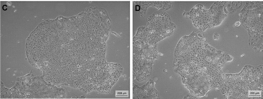 Fig. 1. Phase-contrast microscopic views of CCD841 CoN cells at early and late passages in culture (C and D) (Sariboyaci A E, Uysal O, et al., 2022).
5-Keto PGE2 Upregulates Expression of HO-1 in CCD 841 CoN Cells
Prostaglandin E2 (PGE2) overproduction caused by COX-2 overexpression is characteristic of inflammatory bowel disease (IBD) and inflammation-associated cancer. 15-hydroxyprostaglandin dehydrogenase (15-PGDH) inactivates PGE2 to 15-keto PGE2, and 15-PGDH is a tumor suppressor. In this study, Lee et al. used human colon epithelial CCD 841 CoN cells to determine whether 15-keto PGE2 activated Nrf2 signaling, leading to HO-1 expression.
They determined the non-cytotoxic concentrations of 15-keto-PGE2 (20 μM and 40 μM) in human colon epithelial CCD 841 CoN cells by MTT assay. These concentrations were not cytotoxic up to 24 h. They found that 15-keto-PGE2 (40 μM) induced HO-1 expression, as determined by Western blot (Fig. 1A) and immunocytochemical analysis (Fig. 1B). To check if 15-keto-PGE2 could induce other antioxidant enzymes like TXN and NQO1, CCD 841 CoN cells were treated for 3, 6, 12, and 24 h. HO-1 mRNA and protein levels were increased at 6 h and maintained up to 12 h, and TXN and NQO1 expression was not induced (Fig. 1C and D).
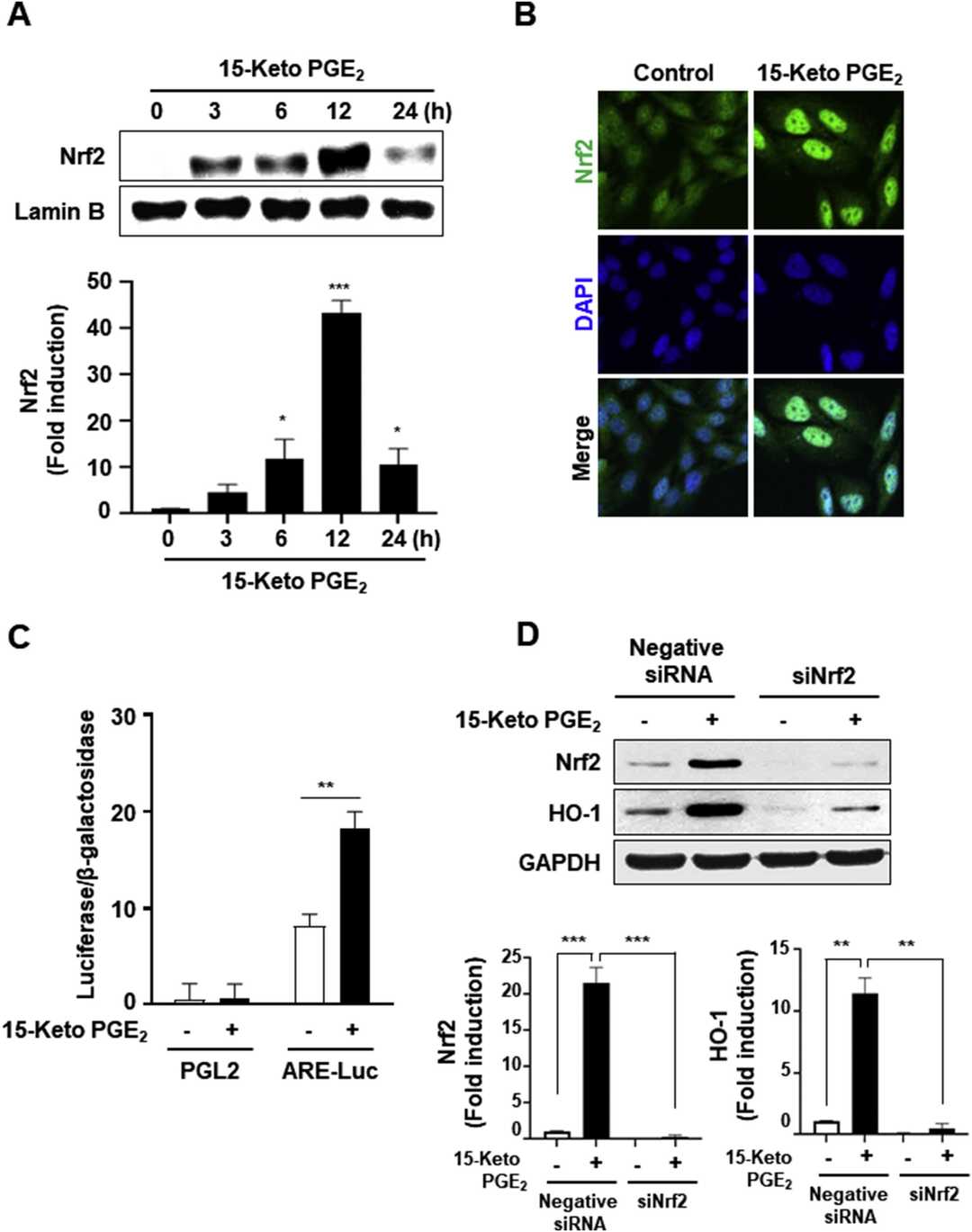 Fig. 1. 15-Keto PGE2 induced expression of HO-1 in human colon epithelial CCD 841 CoN cells (Lee J E, Zhong X C, et al., 2020).
Acrolein Activates the RAS/MAPK Signaling Pathway in CRC Tumorigenesis
Colorectal cancer (CRC) is one of the most common cancers with increasing incidence and low survival rate. Dietary habits including a high-fat diet (HFD) increase the incidence of CRC. Acrolein is a Group 2A carcinogen that is produced during food preparation and intake of HFD. Here, Tsai et al. sought to determine if acrolein plays a role in CRC tumorigenesis through the RAS-MAPK pathway.
To elucidate the oncogenic mechanisms of acrolein, researchers conducted cDNA microarray analysis in NIH/3T3 Acr-clone#4 followed by comprehensive pathway analysis (Fig. 2A). Four genes (Rnd1, Rras2, myc, and PI3Kcb) in the RAS/MAPK pathway were significantly upregulated in acrolein-transformed clone #4 (Fig. 2B). These results were confirmed by Western blot analysis (Fig. 2C). Acrolein activated the RAS/MAPK pathway and induced c-myc in NIH/3T3 cells and human colon epithelium CCD-841CoN (Fig. 2D and E). Acrolein also induced cell proliferation, colony formation, and migration in CCD-841CoN cells (Fig. 3A and D) and activated the RAS/MAPK pathway in CCD-841CoN Acr clone and human colon cancer cell lines SW480 and HCT116 (Fig. 3E). Taken together, these data demonstrate that acrolein induces oncogenic transformation through the RAS/MAPK pathway.
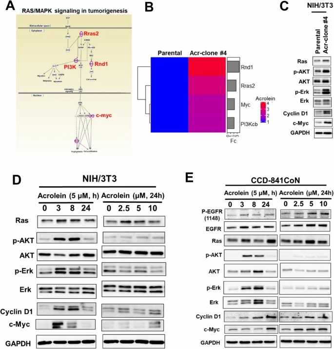 Pathway analysis of gene expression profiles in acrolein-transformed clones (Tsai H C, Tsou H H, et al., 2021).
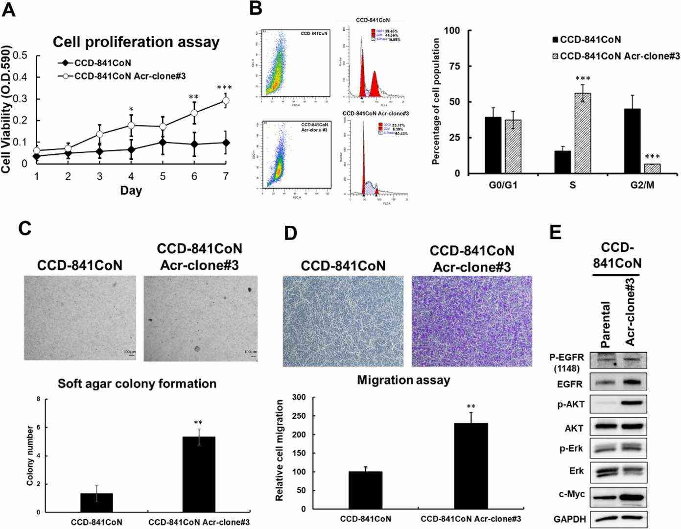 Fig. 3. Acrolein induced oncogenic transformation in CCD-841CoN cells. CCD-841CoN cells were treated with acrolein (Acr, 7.5 μM) for one month and named CCD-841CoN Acr-clone#3 (Tsai H C, Tsou H H, et al., 2021).
Ask a Question
Write your own review
- You May Also Need

Description: Established from an adenocarcinoma located in the ascending colon of a 68 year-old male patient. The adenocarcinoma was well differentiated and determined to be Dukes' stage B. Imperial College ...

Description: This is one cell line out of a series of colon carcinoma cell lines established by PD Dr. Michael Linnebacher.
Description: Rectal carcinoma from a Japanese patient. Cell growth is slow.
Description: The C80 cell line was established from a 69-year old male patient with a moderately well differentiated adenocarcinoma of the rectum classified as Dukes' stage D.

Description: RKO is a poorly differentiated colon carcinoma cell line developed by Michael Brattain.
- Adipose Tissue-Derived Stem Cells
- Human Neurons
- Mouse Probe
- Whole Chromosome Painting Probes
- Hepatic Cells
- Renal Cells
- In Vitro ADME Kits
- Tissue Microarray
- Tissue Blocks
- Tissue Sections
- FFPE Cell Pellet
- Probe
- Centromere Probes
- Telomere Probes
- Satellite Enumeration Probes
- Subtelomere Specific Probes
- Bacterial Probes
- ISH/FISH Probes
- Exosome Isolation Kit
- Human Adult Stem Cells
- Mouse Stem Cells
- iPSCs
- Mouse Embryonic Stem Cells
- iPSC Differentiation Kits
- Mesenchymal Stem Cells
- Immortalized Human Cells
- Immortalized Murine Cells
- Cell Immortalization Kit
- Adipose Cells
- Cardiac Cells
- Dermal Cells
- Epidermal Cells
- Peripheral Blood Mononuclear Cells
- Umbilical Cord Cells
- Monkey Primary Cells
- Mouse Primary Cells
- Breast Tumor Cells
- Colorectal Tumor Cells
- Esophageal Tumor Cells
- Lung Tumor Cells
- Leukemia/Lymphoma/Myeloma Cells
- Ovarian Tumor Cells
- Pancreatic Tumor Cells
- Mouse Tumor Cells