Sheep Bone Marrow-derived Mesenchymal Stem Cells
- Specification
- Background
- Scientific Data
- Q & A
- Customer Review
Sheep bone marrow-derived mesenchymal stem cells (Sheep BMSCs) are a component of the bone marrow stroma and are one of the non-hematopoietic cell populations in the bone marrow. BMSCs are important for the maintenance of hematopoiesis and the homeostasis of the bone marrow microenvironment. BMSCs have self-renewal and multipotent differentiation potential, and can differentiate into different mesenchymal lineages, including bone, cartilage, adipose, tendon, muscle, and BMSCs themselves.
Sheep BMSCs exhibit strong proliferative capacity and can be expanded in vitro over multiple passages. However, as the number of passages increases, the proliferation rate of Sheep BMSCs gradually decreases, which may be due to cellular senescence. Studies have shown that Sheep BMSCs maintain a stable phenotype during passaging and are able to preserve their undifferentiated state under standard culture conditions. In addition, some studies have found that telomerase reverse transcriptase (TERT) can improve the proliferative capacity of Sheep BMSCs and extend their in vitro lifespan. Sheep BMSCs are commonly used in tissue engineering studies, such as the regeneration of cartilage, bone, and skin tissue.
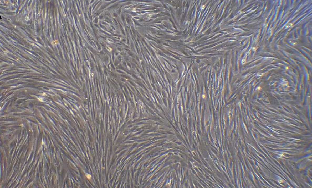 Fig. 1. Morphology of sheep bone marrow-derived mesenchymal stem cell at passage 3 (Zhu X, Zhou L, et al., 2020).
Morphology of Ovine Bone Marrow Derived Mesenchymal Stem Cells Stimulated with FGF-2 and BMP-2
Bone defects and bone disorders are a great challenge for clinicians due to the limits of traditional bone grafts. Mesenchymal stem cells (MSCs) have great potential as cell source in bone regeneration due to their self-renewal and multi-differentiation potential. The purpose of Gromolak et al. is to study the biological properties and osteogenic differentiation potential of ovine bone marrow-derived MSCs (BM-MSCs) after FGF-2 and BMP-2 stimulation to get a better understanding of this material as a potential cellular therapy for bone regeneration in large animal models.
The morphology of BM-MSCs after 21 days culture in different media: (1) complete αMEM, (2) αMEM with FGF-2, (3) αMEM with FGF-2 and BMP-2, (4) osteogenic differentiation medium, (5) osteogenic differentiation medium with FGF-2 and (6) osteogenic differentiation medium with FGF-2 and BMP-2. BM-MSCs grown in αMEM without any additional cytokines maintained the typical spindle shape (Fig. 1a). The cells cultured in αMEM with FGF-2 were smaller and denser, still spindle-shaped (Fig. 1c). The largest changes were noted in a group treated with both BMP-2 and FGF-2, where some cells formed mesh-like structures and some were aggregated to rounded bone-like shapes (Fig. 1e). Calcium deposition was present in all osteogenic differentiation media, where crystals were smaller and more dispersed in media supplemented with BMP-2 and/or FGF-2 (Fig. 1b,d,f).
 Fig. 1. Morphological changes of sheep bone marrow-derived mesenchymal stem cells in different culture conditions (Gromolak S, Krawczenko A, et al., 2020).
Telomerase Enhances Osteogenic Differentiation of Sheep Bone Marrow Mesenchymal Stem Cells (BMSCs) by Up-Regulating PI3K/Akt Pathway in vitro
Bone marrow mesenchymal stem cells (BMSCs) have the multi-directional differentiation and are widely used in bone tissue engineering. After long-term in vitro culture, BMSCs are prone to aging and differentiation loss with multiple passages. Telomerase reverse transcriptase (TERT) can extend telomeres, thus supporting cell proliferation and differentiation.
In this study, TERT vectors were transfected into sheep BMSCs to construct TERT-BMSC cell lines. Transcriptome sequencing, RT-qPCR, and Western blot were used to explore the influence of ectopic expression of TERT on osteogenic differentiation and the role of PI3K/Akt pathway. The BMSCs and TERT-BMSCs remain in spindle shape at passage 3 (Figs. 2A, 2B). Passage 7 BMSCs is flat, large and edge blurred (Fig. 2C). The passage 30 TERT-BMSC is still in the original shape and has strong proliferation (Fig. 2D). Even at >7 months (>45 passages) (Fig. 2E), the TERT-BMSCs are not obvious senescence. Transcriptome sequencing showed that cyclins (CCNA2, CCNB1, CCND1, CCND2, CCNE1, CCNE2), CDKs (CDK1, CDK6), RBL1 and E2F family members (E2F1, E2F2) are significantly upregulated in TERT-BMSCs compared to BMSCs. Cyclins (CCNA1, CCND3) and CDKs (CDK2, CDK4) show no significant difference between the two. CDKN1A/p21 and CDKN2A/p16 are significantly higher, while CDKN1B/p27 is significantly lower in TERT-BMSCs than in BMSCs.
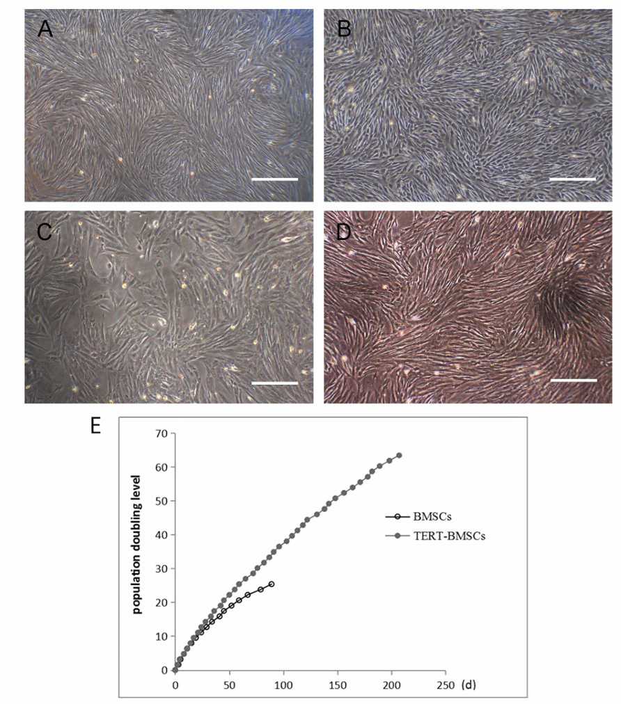 Fig. 2. Comparison of morphology and proliferation between BMSCs and TERT-BMSCs (Zhu X, Zhou L, et al., 2020).
Ask a Question
Write your own review
- Adipose Tissue-Derived Stem Cells
- Human Neurons
- Mouse Probe
- Whole Chromosome Painting Probes
- Hepatic Cells
- Renal Cells
- In Vitro ADME Kits
- Tissue Microarray
- Tissue Blocks
- Tissue Sections
- FFPE Cell Pellet
- Probe
- Centromere Probes
- Telomere Probes
- Satellite Enumeration Probes
- Subtelomere Specific Probes
- Bacterial Probes
- ISH/FISH Probes
- Exosome Isolation Kit
- Human Adult Stem Cells
- Mouse Stem Cells
- iPSCs
- Mouse Embryonic Stem Cells
- iPSC Differentiation Kits
- Mesenchymal Stem Cells
- Immortalized Human Cells
- Immortalized Murine Cells
- Cell Immortalization Kit
- Adipose Cells
- Cardiac Cells
- Dermal Cells
- Epidermal Cells
- Peripheral Blood Mononuclear Cells
- Umbilical Cord Cells
- Monkey Primary Cells
- Mouse Primary Cells
- Breast Tumor Cells
- Colorectal Tumor Cells
- Esophageal Tumor Cells
- Lung Tumor Cells
- Leukemia/Lymphoma/Myeloma Cells
- Ovarian Tumor Cells
- Pancreatic Tumor Cells
- Mouse Tumor Cells