DH82
Cat.No.: CSC-C9176W
Species: Canis lupus familiaris (Dog)
Morphology: macrophage
- Specification
- Background
- Scientific Data
- Q & A
- Customer Review
The DH82 cell line was developed from neoplastic tissue of a 10-year-old male Golden Retriever with malignant histiocytosis which demonstrates typical macrophage characteristics. While the DH82 cell line expresses the Fcγ receptor it lacks expression of the Fcμ and C3b receptors and fails to produce interleukin-1 (IL-1). The DH82 cells secrete several cytokines and chemokines including TNF-α and IL-6 while expressing P2Y2 receptors yet show limited intracellular P2X4 receptor expression.
The DH82 cell line demonstrates extensive phagocytic capabilities when active in immune processes. The microbicidal function of this cell line gets blocked by capsular polysaccharides from Cryptococcus neoformans including GXMGx and GXM. This model stands out as an essential framework for studying how macrophages interact with pathogens. Researchers utilize the DH82 cell line as a primary research tool in canine medical science to study canine macrophage responses to infections through changes in signaling pathways and gene expression regulation.
Scientists use this cell line to explore how canine histiocytic sarcoma develops and what treatment methods can be effective. CDV infection triggers reduced anti-angiogenic gene expression in DH82 cells which results in tumor growth suppression. The study results provide theoretical support for developing novel treatment strategies against histiocytic sarcoma.
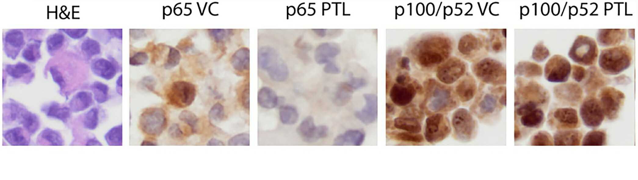 Fig. 1. Hematoxylin and eosin (left-most panel) and p65 and p52 immunohistochemistry (remaining panels) of canine cell line cell pellets treated with vehicle control (VC) or the nuclear factor kappa B (NF-kB) inhibitor parthenolide (PTL) (Schlein LJ, Thamm DH, et al., 2023).
Purified Polysaccharides from C. neoformans Inhibit Binding and Phagocytic Activity of Canine Macrophages DH82
Cryptococcus neoformans is a harmful fungus affecting both the respiratory and central nervous systems. The macrophage is a crucial cell type in the immune response against this infection, but these cells can be modulated by the fungus's capsular polysaccharides, GXM and GXMGal, enabling the pathogen to evade immune detection. LaRocque-de-Freitas et al. aimed to examine if these polysaccharides impact the microbicidal functions of canine macrophages, specifically focusing on DH82 cell lines.
For this purpose, DH82 macrophage cell cultures were established and treated with GXM-Gal, GXM, or C. neoformans total capsule at different concentrations, followed by a 24 h incubation period. Apoptotic cells were measured using annexin V/PI staining. Results showed no apoptosis induction in DH82 cells by polysaccharides or total capsule (Fig. 1). Macrophages' pathogen binding and phagocytosis are vital for eliminating infections. They examined the effect of GXM-Gal, GXM, or total capsule on DH82 cell binding and phagocytosis. DH82 cells incubated with polysaccharides or total capsule showed reduced yeast binding and phagocytic activity. Fewer yeasts were attached to DH82 cells (Fig. 2A), and fewer cells could phagocytose yeasts (Fig. 2B and C). This suggests C. neoformans polysaccharides impair DH82 cells' binding and phagocytosis abilities.
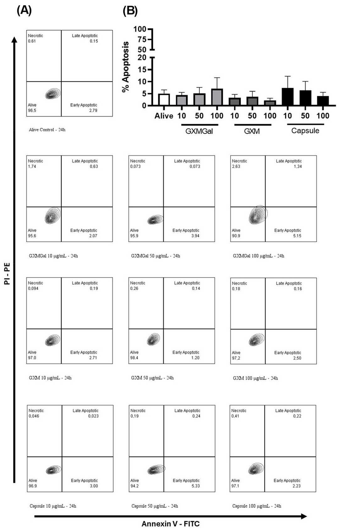 Fig. 1. The effect of purified polysaccharides and the total capsule on the apoptosis induction of DH82 cells (LaRocque-de-Freitas IF, da Silva-Junior EB, et al., 2024).
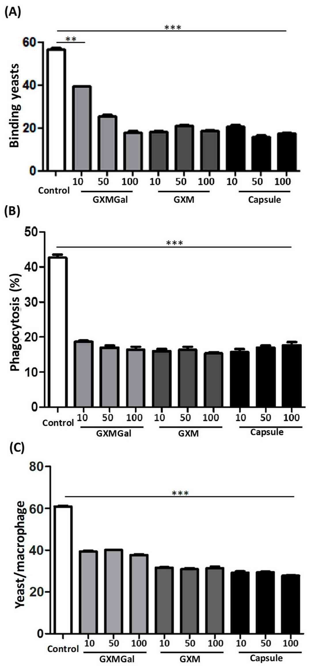 Fig. 2. Binding and phagocytosis by DH82 in the presence of capsular polysaccharides of C. neoformans (LaRocque-de-Freitas IF, da Silva-Junior EB, et al., 2024).
Infectivity of Aflagellar Epimastigotes of Trypanosoma caninum in the DH82 Cell Line and Mouse Peritoneal Macrophages
Trypanosoma caninum infects domestic dogs, especially in areas prone to leishmaniasis. It appears in various forms in cultures, but previous attempts to induce infection in models have failed. This study used in vitro interactions between aflagellar epimastigotes of T. caninum, DH82 cells, and mouse macrophages, utilizing microscopy techniques to observe interactions and transformations.
Using BF microscopy, they detected T. caninum adhering to macrophages within 15 minutes, alongside free amastigotes and epimastigote interactions, with noted vacuoles in macrophages. SEM revealed multiple adhesion patterns of aflagellar epimastigotes to DH82 cells, including body-wide adhesion (Fig. 3a). At 30 and 45 minutes, extracellular amastigotes formed clusters interacting with macrophages (Fig. 3b). SEM and TEM showed amastigote-macrophage interactions and structural details, including shape changes (Fig. 3c). Two hours in, free aflagellar epimastigotes interacted with DH82 cells (Fig. 3d). At 6 hours, amastigote interaction caused cell disruption, with TEM revealing detailed amastigote structures (Fig. 3e). By 24 hours, amastigote clusters appeared to fracture the cells (Fig. 3f), and shape transition was observed (Fig. 3g). At 48 hours, free amastigotes and macrophage interactions continued (Fig. 3h).
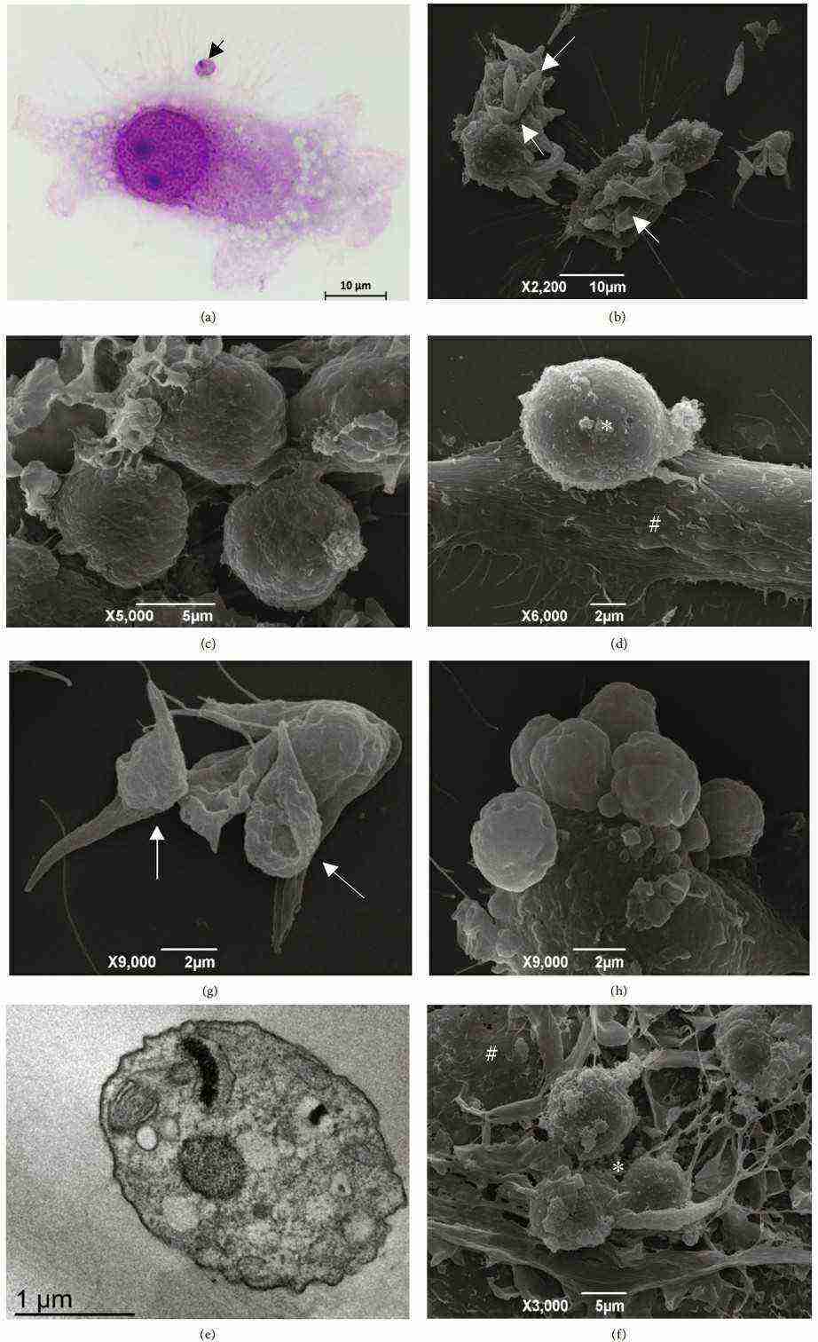 Fig. 3. DH82 cell interactions with T. caninum (Nascimento KCS, de Oliveira Souza SM, et al., 2025).
Ask a Question
Write your own review
- You May Also Need
Description: MDCK II - Madin-Darby canine kidney- is a spontaneously immortalized cell line subcloned from the heterogenous parent line - MDCK.
- Adipose Tissue-Derived Stem Cells
- Human Neurons
- Mouse Probe
- Whole Chromosome Painting Probes
- Hepatic Cells
- Renal Cells
- In Vitro ADME Kits
- Tissue Microarray
- Tissue Blocks
- Tissue Sections
- FFPE Cell Pellet
- Probe
- Centromere Probes
- Telomere Probes
- Satellite Enumeration Probes
- Subtelomere Specific Probes
- Bacterial Probes
- ISH/FISH Probes
- Exosome Isolation Kit
- Human Adult Stem Cells
- Mouse Stem Cells
- iPSCs
- Mouse Embryonic Stem Cells
- iPSC Differentiation Kits
- Mesenchymal Stem Cells
- Immortalized Human Cells
- Immortalized Murine Cells
- Cell Immortalization Kit
- Adipose Cells
- Cardiac Cells
- Dermal Cells
- Epidermal Cells
- Peripheral Blood Mononuclear Cells
- Umbilical Cord Cells
- Monkey Primary Cells
- Mouse Primary Cells
- Breast Tumor Cells
- Colorectal Tumor Cells
- Esophageal Tumor Cells
- Lung Tumor Cells
- Leukemia/Lymphoma/Myeloma Cells
- Ovarian Tumor Cells
- Pancreatic Tumor Cells
- Mouse Tumor Cells