
Rat Tumor Cells
Rat tumor cell lines are a well-established model system in oncology, toxicology, neurobiology and pharmacology research due to their stable growth features and high compatibility with rat illness models. These models are essential tools for researching tumor formation, carcinogenesis, signaling pathways, treatment response and tumor-host interactions.
Our rat tumor cell bank comprises a broad spectrum of frequently used cell models from glioma, hepatoma, pituitary tumor, mammary tumor, adrenal tumor and other tumors. Representative cell lines such as C6, RG2, GH3, INS-1, N1-S1 and Walker 256 enable numerous applications including cancer biology, drug screening, endocrine research, neuro-oncology and metabolic illness studies.
Well-Characterized Experimentally Versatile Research-Validated Supported
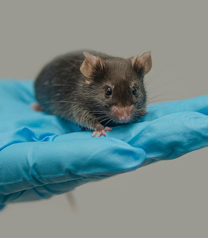
Key Features & Expertise
Our rat tumor cell models support a wide range of oncology and translational research applications

Diverse Rat Tumor Models
- Includes widely used glioma, hepatoma, endocrine, and mammary tumor cell models
- Suitable for studies of proliferation, invasion, angiogenesis, and metastasis
- Support investigations into cancer signaling and disease progression mechanisms

Widely Applied in Translational Research
- Compatible with pharmacology, toxicology, and neuro-oncology studies
- Frequently used in endocrine function and metabolic disease research
- Suitable for drug screening, imaging, and pathway analysis workflows

Reliable Quality & Experimental Consistency
- Quality controlled for stable and reproducible experimental performance
- Mycoplasma-free and maintained using standardized culture conditions
- Cryopreserved stocks with technical support and stable supply available
FAQ
Are rat tumor cells suitable for neuro-oncology studies?
Yes. Rat glioma models such as C6 and RG2 are frequently utilised for investigating brain tumor biology, tumor invasion, angiogenesis and therapeutic response. They are also commonly employed in orthotopic animal models of glioma research.
Can rat tumor cell lines be used in endocrine and metabolic research?
Of course. Some rat tumor-derived models, such as GH3 and INS-1 cells, are widely employed in the investigations of endocrine signalling, hormone secretion, pancreatic beta-cell function, diabetes and metabolic disorders.
Are these cell lines suitable for drug screening applications?
Yes. Rat tumor cells are commonly employed in cytotoxicity test, pathway inhibition research, toxicological assessment and preclinical drug screening workflows because of their stable growth and predictable responses.
How are rat tumor cells shipped and stored?
Cells are typically supplied as cryopreserved vials and shipped on dry ice. For long-term preservation and optimal viability, storage in liquid nitrogen is recommended after receipt.
Filters Clear all filters
Species
- African clawed frog (1)
- American mink (1)
- Asian tiger mosquito (1)
- Atlantic salmon (1)
- Bluegill (2)
- Bluestriped grunt (1)
- Bovine (7)
- Brazilian free-tailed bat (1)
- Brown bullhead (2)
- Cabbage looper (1)
- Cabbage moth (6)
- Cat (3)
- Central mudminnow (1)
- Chicken (3)
- Chinese hamster (5)
- Chinook salmon (2)
- Chum salmon (1)
- Coho salmon (1)
- Common carp (2)
- Cotton-top tamarin (1)
- Dog (2)
- Fall armyworm (3)
- Fathead minnow (2)
- Fruit fly (1)
- Gilthead sea bream (2)
- Golden hamster (7)
- Goldfish (6)
- Gray dwarf hamster (1)
- Green monkey (2)
- Gypsy moth (1)
- Horse (1)
- Japanese eel (1)
- Japanese rice fish (7)
- Koi carp (1)
- Mouse (310)
- Mouse x Gray dwarf hamster (1)
- Mouse x Rat (20)
- Northern pike (1)
- Pig (3)
- Rabbit (2)
- Rainbow trout (3)
- Rat (114)
- Rhesus macaque (1)
- Salt marsh moth (1)
- Sheep (2)
- Snakehead murrel (2)
- Sockeye salmon (1)
- Vervet monkey (2)
- Zebrafish (2)
Source
- Abdomen (1)
- Adipose (2)
- Adrenal Gland (1)
- Aorta (4)
- Artery (1)
- Ascites (5)
- Ascites Metastasis (5)
- Bladder (11)
- Bladder Metastasis (1)
- Blastocyst (1)
- Blastula (1)
- Blood (7)
- Bone (6)
- Bone Marrow (14)
- Brain (24)
- Brain Metastasis (1)
- Breast (22)
- Caudal Peduncle (1)
- Caudal Trunk (2)
- Colon (6)
- Connective Tissue (7)
- Dermis (1)
- Embryo (29)
- Fetus (2)
- Fin (9)
- Glomerulus (2)
- Head Kidney (2)
- Heart (4)
- Hemolymph (1)
- Ileum (1)
- Intestine (9)
- Jejunum (1)
- Kidney (18)
- Liver (22)
- Lung (16)
- Lymph Node (2)
- Lymph Node Metastasis (1)
- Muscle (3)
- Ovary (8)
- Pancreas (9)
- Peripheral Blood (7)
- Peripheral Nervous System (21)
- Pituitary Gland (7)
- Prostate (3)
- Rectum (2)
- Skeletal Muscle (4)
- Skin (10)
- Small Intestine (3)
- Smooth Muscle (2)
- Soft Tissue (1)
- Spinal Cord (2)
- Testis (6)
- Thymus (5)
- Thyroid Gland (1)
- Trachea (1)
- Uterus (1)
Disease
- Bovine Leukemia (2)
- Canine Histiocytic Sarcoma (1)
- Chicken Bursal Lymphoma (2)
- Goldfish Erythrophoroma (4)
- Hamster Kidney Tumor (1)
- Hamster Pancreatic Ductal Adenocarcinoma (1)
- Hamster Uterine Leiomyosarcoma (1)
- Medaka Hepatoma (2)
- Mouse Bladder Transitional Cell Carcinoma (1)
- Mouse Chondrosarcoma (1)
- Mouse Colon Adenocarcinoma (3)
- Mouse Ependymoma (2)
- Mouse Erythroid Leukemia (13)
- Mouse Fibrosarcoma (5)
- Mouse Glioblastoma (1)
- Mouse Hemangioendothelioma (1)
- Mouse Hepatocellular Carcinoma (1)
- Mouse Insulinoma (3)
- Mouse Islet Cell Adenoma (1)
- Mouse Kidney Carcinoma (1)
- Mouse Leukemia (10)
- Mouse Leydig Cell Tumor (1)
- Mouse Lymphoma (8)
- Mouse Mammary Gland Malignant Neoplasm (21)
- Mouse Melanoma (9)
- Mouse Multiple Myeloma (5)
- Mouse Myeloid Leukemia (3)
- Mouse Neoplasm (1)
- Mouse Neuroblastoma (21)
- Mouse Oral Cavity Squamous Cell Carcinoma (1)
- Mouse Osteosarcoma (3)
- Mouse Pituitary Gland Neoplasm (1)
- Mouse Precursor T Cell Lymphoblastic Lymphoma/Leukemia (2)
- Mouse Pulmonary Adenoma (1)
- Mouse Pulmonary Malignant Tumor (3)
- Mouse Pulmonary Squamous Cell Carcinoma (1)
- Mouse Rectum Carcinoma (2)
- Mouse Reticulum Cell Sarcoma (2)
- Mouse Sarcoma (1)
- Mouse Teratocarcinoma (8)
- Mouse Thymic Lymphoma (3)
- Rat C-Cell Carcinoma (1)
- Rat Cholangiocarcinoma (1)
- Rat Colon Adenocarcinoma (5)
- Rat Digestive System Neoplasm (1)
- Rat Fibrosarcoma (1)
- Rat Hepatocellular Carcinoma (20)
- Rat Histiocytic Sarcoma (1)
- Rat Insulinoma (2)
- Rat Leukemia (1)
- Rat Leydig Cell Adenoma (1)
- Rat Lung Carcinoma (1)
- Rat Malignant Glioma (4)
- Rat Malignant Meningioma (1)
- Rat Malignant Oligodendroglioma (2)
- Rat Malignant Thymoma (3)
- Rat Mammary Gland Adenocarcinoma (10)
- Rat Neuroblastoma (3)
- Rat Osteosarcoma (2)
- Rat Pituitary Gland Neoplasm (6)
- Rat Prostate Adenocarcinoma (3)
- Rat Rhabdomyosarcoma (1)
- Rat Sarcoma (2)
- Rat Squamous Cell Carcinoma (1)
- Rat Urinary Bladder Transitional Cell Carcinoma (2)
- Rat Urinary System Neoplasm (6)
Description: Established ca. 1986 from the liver of a diethylnitrosamine-treated male Wistar AF/Han rat
Description: Established ca. 1986 from the liver of a diethylnitrosamine-treated male Wistar AF/Han rat
Description: Established ca. 1986 from the liver of a diethylnitrosamine-treated male Wistar AF/Han rat
Description: Established ca. 1986 from the liver of a diethylnitrosamine-treated male Wistar AF/Han rat
Description: Established from a spontaneously developed pancreatic carcinoma of a Wistar AF/Han rat; cells were ...
Description: Established ca. 1986 from the hepatocellular carcinoma induced in vivo by chronic nutritional ...
Description: Established from a spontaneously developed rhabdomyosarcoma of a Wistar AF/Han rat
Description: Established ca. 1986 from the cholangio-hepatocellular tumor induced in vivo by treatment of a male ...

Description: Sertoli cells dissected from normal testis from 17-day-old Wistar rat and immortalized with SV40 ...
Description: Established from the thoracic aorta of a DB1X embryonic rat in 1976; cells were described to ...
Description: Species: rat (Rattus norvegicus)Cell type: kidney epithelial-like cellsOrigin: cell line was cloned ...
Description: Species: rat; Tissue: liver; Tumor: hepatoma, H-35 Reuber; Derived from: H4-II-E-C3
Description: Species: rat, buffalo male; Tissue: ascitic fluid; Tumor: hepatoma; Derived from: Morris hepatoma ...
Description: Species: rat, Lewis; Tumor: fibrosarcoma; Transformed by: RSV-33; tumor induced in vivo by RSV-33; ...
Description: Established from the normal liver of a 17-day rat embryo (strain JAR-2) in 1975

Description: Established from isolated renal glomeruli of a 3-week-old normal male Wistar rat in 1988
Description: The AY-27 cell line was established from a primary bladder tumor induced by ...
Description: Normal rat epithelial cells from the small intestine; cells were described to synthesize ...