mouse bone marrow mesenchymal stem cells (B129 MBMMSC)
- Specification
- Background
- Scientific Data
- Q & A
- Customer Review
Each batch of Mouse Bone Marrow Mesenchymal Stem Cells are tested for expression of markers using antibodies, CD44, Sca-1 and CD29 by flow cytometry.
Mouse Bone Marrow Mesenchymal Stem Cells (B129 MBMMSC) are primary bone marrow-derived mesenchymal stem/stromal cells (MSCs) derived from the bone marrow of B129 mice. They are a population of multipotent adult stem cells that can self-renew and differentiate into multiple cell lineages and have been used as a tool for many areas of regenerative medicine, immunology, and musculoskeletal-related studies in vitro.
Like most MSCs, B129 MBMMSCs are fibroblast-like, exhibit a spindle-shaped morphology, and adhere to conventional tissue culture plastic. They have also been shown to express mesenchymal markers including CD29, CD44, CD73, CD90 and Sca-1 and lack expression of hematopoietic and endothelial markers such as CD34, CD45 and CD11b. B129 MBMMSCs also maintain functionality and can differentiate into osteogenic, adipogenic and chondrogenic lineages with proper induction (defined by alkaline phosphatase staining and mineralization, Oil Red O staining, and collagen production respectively).
As these cells are derived from mice and can easily be obtained from mice that are genetically engineered to study disease progression, B129 MBMMSCs can be used to bridge in vitro studies to in vivo disease models. They can be used to study bone regeneration, cartilage repair, hematopoietic niche formation and function, fibrosis, immune system interaction, and inflammatory signaling. B129 MBMMSCs have been used as a tool to screen biomaterials, scaffolds, and gene modifications.
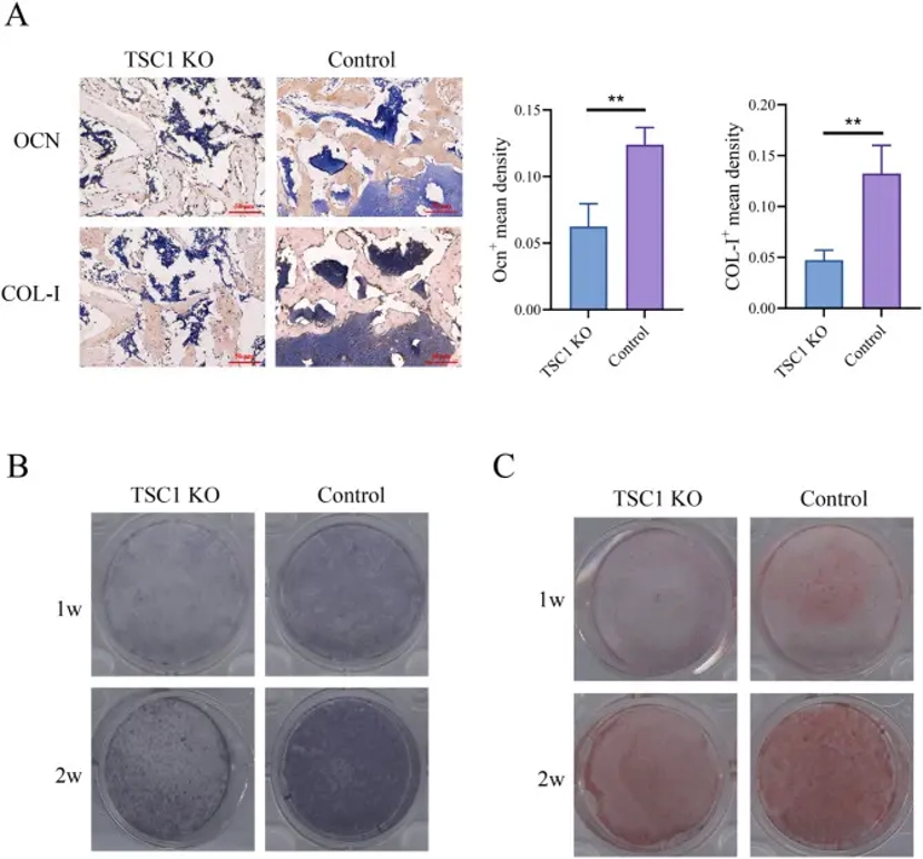
mTORC1 Activation Inhibits the Differentiation of BMMSCs into Osteoblasts
Bone defects from trauma and orthopedic diseases present major clinical challenges due to treatment difficulty, duration, and cost. Huang's team demonstrated that activated mTOR signaling in mesenchymal stromal cells regulates bone repair.
During repair, bone marrow MSCs (BMMSCs) recruit to injury sites, undergo osteogenic differentiation, and contribute to callus formation through ECM secretion. To assess mTORC1 activation effects, they examined tibial sections from TSC1 KO and control mice at 2 weeks post-injury. TSC1 KO showed markedly downregulated osteogenic markers OCN and collagen I, with reduced osteoblast numbers (P = 0.001), indicating impaired in vivo osteogenesis (Fig. 1A). In vitro validation using isolated MSC progenitors confirmed these findings: TSC1 KO-derived BMMSCs showed significantly weaker mineral deposition and ALP activity at 1 and 2 weeks of osteogenic induction versus controls (Fig. 1B, C), demonstrating consistent osteogenic defects due to mTORC1 activation.
Ask a Question
Write your own review
- You May Also Need
- Adipose Tissue-Derived Stem Cells
- Human Neurons
- Mouse Probe
- Whole Chromosome Painting Probes
- Hepatic Cells
- Renal Cells
- In Vitro ADME Kits
- Tissue Microarray
- Tissue Blocks
- Tissue Sections
- FFPE Cell Pellet
- Probe
- Centromere Probes
- Telomere Probes
- Satellite Enumeration Probes
- Subtelomere Specific Probes
- Bacterial Probes
- ISH/FISH Probes
- Exosome Isolation Kit
- Human Adult Stem Cells
- Mouse Stem Cells
- iPSCs
- Mouse Embryonic Stem Cells
- iPSC Differentiation Kits
- Mesenchymal Stem Cells
- Immortalized Human Cells
- Immortalized Murine Cells
- Cell Immortalization Kit
- Adipose Cells
- Cardiac Cells
- Dermal Cells
- Epidermal Cells
- Peripheral Blood Mononuclear Cells
- Umbilical Cord Cells
- Monkey Primary Cells
- Mouse Primary Cells
- Breast Tumor Cells
- Colorectal Tumor Cells
- Esophageal Tumor Cells
- Lung Tumor Cells
- Leukemia/Lymphoma/Myeloma Cells
- Ovarian Tumor Cells
- Pancreatic Tumor Cells
- Mouse Tumor Cells