Primary Cultures of HUVECs
Introduction
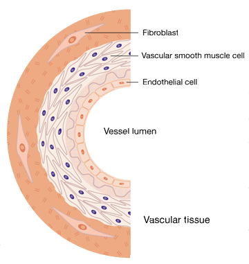
Endothelium, once viewed as a passive layer of cells forming a barrier between the blood and tissues, is now considered to act as a distributed organ, whose functions are critical for normal homeostasis. Endothelial cells line is on the lumina of all blood vessels. Because of their unique position, they are the only cells in the body that form an interface among a fluid moving under relatively high pressure, the blood, and a solid substrate, the vessel wall. The endothelium is involved in many critical processes such as maintaining vascular tone, acting as a selectively permeable barrier, regulating coagulation and thrombosis, and directing the passage of leukocytes into areas of inflammation.
In order to gain insight into the role of endothelial cells in normal and pathological states, human umbilical vein endothelial cells were isolated from the endothelium of veins from the newborn's umbilical cord. HUVECs have been already used extensively to study the biology and pathobiology of the human endothelial cell, and most of our knowledge of human endothelial cells is derived from experiments with cultured HUVECs.
The main advantage of using HUVECs to study endothelial biology is the wide availability of the umbilical cord, a relatively simple method of isolation, and the general purity of the cell population obtained.
At Creative Bioarray, we provide primary HUVECs and complete medium. In addition, HUVECs characterization services are also available. If you have any other requirements, please feel free to contact us.
Here, we will describe the basic procedures of HUVECs culture (including cryopreservation) and the methods used to characterize the phenotype of endothelial cells.
Materials
1. Phosphate-buffered saline (PBS).
2. Fetal bovine serum (FBS).
3. HUVEC culture medium.
Notes:
It is advisable to aliquot HUVEC medium in 50-mL tubes that are kept at 4°C until use to avoid repeated warming and
cooling of medium.
4. Trypsin EDTA. A 5X stock solution of trypsin EDTA is prepared as below table.
| Component | Volume added for 200 mL | Final concentration |
|---|---|---|
| Trypsin | 2.5 g | 0.25% |
| EDTA (250 mM) | 4 mL | 1 mM |
| PBS (10X) | 100 mL | 1X |
| Deionized water | To final volume of 200 mL |
Notes: The solution is sterilized by filtration through a 0.2-µm filter. Dispense 10-mL aliquots
into 50-mL tubes and store at –20°C. Before use, add 40 mL of sterile deionized water to dilute to 1X. Keep at 4°C
for up to 4 weeks.
5. 50-mL Tubes.
6. 100-mm Tissue culture plates.
7. HUVEC freezing medium. Prepare
fresh as needed by mixing the necessary volumes of 45% FBS, 45% HUVEC medium, and 10% dimethyl sulfoxide (DMSO). The
solution must be ice cold prior to use.
• Subculture of Primary Endothelial Cells
This procedure allows efficient amplification of endothelial cells from primary cultures of human umbilical vein at a relatively low passage number. Continued passage will eventually result in senescent changes and loss of useful replicative potential. Although there is variability among batches of cells, HUVECs can routinely be used for up to 8–10 passages and should be passaged before the cells reach confluence and prior to the growth medium becoming acidic. PBS, trypsin–EDTA, and HUVEC medium should be warmed up at room temperature or 37°C before use.
- Aspirate the medium from a dish of primary HUVECs (isolated from one or more umbilical cords) at or near confluence.
- Wash the monolayer with 5 mL of PBS for a 100-mm dish (use 2 mL for a 60-mm dish).
- Add 2 mL of trypsin–EDTA in PBS, allowing the surface to be covered.
- Incubate the cells at 37°C for 1–3 min. Examine the dish under a microscope to determine that the cells have detached. If cells are rounded but not detached, they can be dislodged by gently tapping the plate.
- Add 5 mL of complete HUVEC medium directly to the dish. The serum in the medium will quench the activity of the trypsin.
- Distribute the diluted cell suspension into the gelatin-precoated tissue culture dishes.
- Bring the final volume in each dish up to 8–10 mL with complete HUVEC medium.
- Incubate the dishes at 37°C in 5% CO2 + 95% air. Refeed with complete HUVEC medium every 2–3d.
• Cryopreserved and Thawing of HUVECs
1. Cryopreserved
It is advisable to freeze HUVECs that have been passaged as few times as possible in order to ensure an adequate stock of low-passage cells for subsequent experiments.
1.1 Choosing early passage to cryopreservation, such as the second passage, trypsinize as described before.
Resuspend the cells in 10 mL of fresh HUVEC medium and transfer to a 15-mL tube.
1.2 Centrifuge for 5 min at 300g and then resuspend the cell pellet in 1 mL of HUVEC medium.
1.3 To prepare for a cell count and determine cell concentration. The ideal concentration is between 5 × 10⁵
and 1 × 10⁶ cells/mL.
1.4 Fill tube with 5 mL of HUVEC medium and centrifuge for 5 min at 300g.
1.5 Aspirate the majority of the media and briefly chill on ice (1–2 min).
1.6 Resuspend the cell pellet in
ice-cold freezing medium.
1.7 Dispense the cells in 1-mL aliquots into the prechilled labeled cryovials.
1.8 Put the cryovials into a propanol-based freezing device overnight. Then Transfer the cryovials to liquid
nitrogen for long-term storage.
2. Throwing
2.1 Warm HUVEC medium to room temperature or 37°C.
2.2 Remove cryovial from liquid nitrogen and immediately place in a rack in a 37°C water bath.
2.3 Transfer cells to a 15-mL centrifuge tube. Add 10 mL of HUVEC medium, drop by drop.
2.4 Centrifuge for 5 min at 300g and then discard the supernatant.
2.5 Add 8 mL of HUVEC medium, pipet gently up and down to a single-cell suspen- sion, and plate into a 100-mm
dish.
2.6 Incubate at 37°C, changing the medium as required.
Notes: Cell viability can be severely compromised if the procedures for freezing and thawing are not carried out carefully. Placing the cryovials in a propanol-based freezing device ensures that freezing occurs slowly and reduces cell death. Similarly, when thawing the cells, it is important to add the medium slowly, as sudden dilution of DMSO can cause severe osmotic damage.
• Characterization of Primary Endothelial Cells
When you use non-commercial HUVECs, characterization assays are necessary. The most effective means of characterizing endothelial cultures is to examine various properties. Characteristic endothelial markers include von Willebrand factor, CD31, CD34, VE-cadherin, VEGF receptors 1 and 2. In addition to biomarker determination, detecting the uptake of AcLDL, staining with Ulex europaeus lectin type 1, and morphology observation are also the main methods.
1. Assessment of Cell Monolayer
The monolayer of endothelial cells is characterized by a "cobblestone" appearance at confluence.
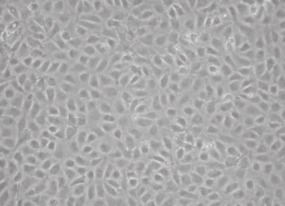
Notes: Morphological identification is not sufficient for the determination of the endothelial phenotype, as endothelial cells can change their morphology depending on the growth supplements in the medium or the matrix onto which the cells are seeded. Indeed, studies have shown that endothelial cells isolated from different organs or different-sized vessels can differ in their antigens, expression of cellular adhesion molecules, metabolism, and growth requirements in culture. It is therefore necessary to use a combination of markers, visual identification, and/or functional assays to confirm the endothelial phenotype.
2. Immunofluorescent Staining of Endothelial Cells
This procedure is used to determine the expression of markers to establish that cells recovered are endothelial. Commercially antibodies are available to detect endothelial markers.
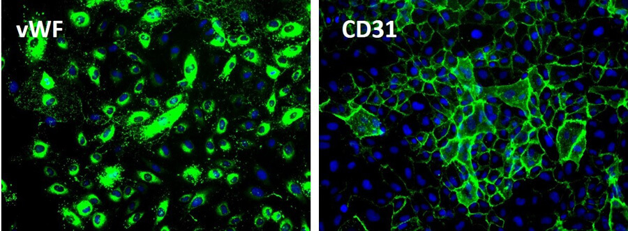
Notes: Most of the markers are not specifically expressed on endothelial cells and that endothelial cell populations show heterogeneous expression of various markers.
3. Uptake of Acetylated LDL
Chemically modified LDL, such as acetylated LDL, are rapidly taken up by macrophages and cultured endothelial cells, and this assay is widely used to isolate and identify endothelial cells in culture. The receptor involved in this pathway is called a scavenger receptor. However, studies have shown that the receptor present on endothelial cells is distinct from the scavenger receptor expressed on macrophages and has been termed scavenger receptor expressed by endothelial cells (SREC). Uptake of acetylated LDL labeled with the fluorescent dye 1,1'-dioctadecyl-3,3,3',3'-tetramethyl indocarbocyanine (DiI) is a fast and convenient way to identify endothelial cells in culture.
Notes: Macrophages will also be labeled by this method.