Featured Products
Our Promise to You
Guaranteed product quality, expert customer support

MHH-CALL-3
- Specification
- Recommended Products
Immunology: CD3 -, CD10 +, CD13 -, CD19 +, CD20 +, CD34 -, CD38 +, cyCD79a +, CD80 -, CD138 +, HLA-DR +
Viruses: ELISA: reverse transcriptase negative; PCR: EBV -, HBV -,
- Background
- Scientific Data
- Q & A
- Customer Review
- Product Information Sheet
The MHH-CALL-3 cell line is a well-known cell line derived from the bone marrow of an 11-year-old girl diagnosed with pre-B cell acute lymphoblastic leukemia (ALL) in 1993. The MHH-CALL-3 cell line represents a specific subtype of pre-B cell ALL and has provided important insights into the disease.
The MHH-CALL-3 cell line has been extensively characterized, including analysis of its immunophenotype, genetic abnormalities, gene expression profiles, and responses to various treatments. These studies have provided valuable information about the molecular pathways involved in pre-B cell ALL and have contributed to the identification of potential therapeutic targets.
Researchers have used the MHH-CALL-3 cell line to study the efficacy of different chemotherapeutic agents, targeted therapies, and immunotherapies in pre-B cell ALL. This cell line has also been utilized to investigate the signaling pathways, genetic mutations, and gene expression patterns associated with the development and progression of the disease.
Bortezomib Induces Apoptosis in Prednisone-Resistant Childhood ALL Cells
The ubiquitin-proteasome degradation pathway (UPDP) is possibly altered and may be involved in multi-agent chemotherapy resistance in childhood ALL patients. The proteasome inhibitor bortezomib blocks proteasomal degradation and hence the UPDP. To investigate whether treatment of ALL with the proteasome inhibitor bortezomib acts synergistically with glucocorticoid treatment, human B-cell precursor leukemic cell lines MHH cALL 2 (PPR) and MHH cALL 3 (PGR) were treated with prednisone (6.2 μM) and various concentrations of bortezomib (1.5-12 nM) for up to 96 hours.
Both cell lines showed a dose-dependent increase in apoptosis after bortezomib single treatment (Fig. 1). Single therapy with 12 nM bortezomib-induced in PGR a 42-fold and in PPR cells a 29-fold higher caspase activity compared to treatment with prednisone alone. However, only the PGR cells showed an additive effect on apoptosis induction after combined treatment with prednisone and bortezomib. In the PPR cell line, the amount of PI-positive, lethal cells increased within 48 hours to 20-fold after combined treatment, using 7 nM bortezomib, compared to single prednisone, and 3-fold compared to single bortezomib treatment. In contrast, apoptosis was reduced compared to bortezomib single treatment.
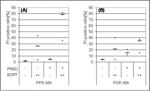 Fig. 1 Increase of PI-positive cells in human B-cell precursor ALL cell lines (MHH cALL 2 and MHH cALL 3). (Junk S, et al., 2009)
Expression of Mer in ALL Cell Lines Correlates with a Quiescent Phenotype in CNS Cocultures
Patients with t(1;19)-positive ALL are prone to central nervous system (CNS) relapses, and expression of the TAM (Tyro3, Axl, and Mer) receptor Mer is upregulated in these leukemias. High Mer expression was detected in MHH-CALL3 and 697 cells with the t(1;19) translocation and REH and UoCB6 cells with the t(12;21) translocation. Low or absent Mer expression was found in SUP-B15 cells carrying the t(9;22) translocation, RS4;11 cells with t(4;11), and in the promyelocytic AML cell line HL-60 (Fig. 2A).
Hypothesizing a role for CNS glia in supporting the survival of Mer-expressing ALL cells, a coculture model with U343 cells was established derived from a low-grade mixed astrocytoma/oligodendroglioma. Coculture of ALL cell lines with U343 for 48 hours resulted in a reduction of viable cells in all cell lines analyzed (Fig. 2B). The observed effect was more pronounced in Merhigh cells and less appreciable in Merlow cells (Fig. 2B). U343 coculture induced a quiescent phenotype in ALL cells characterized by an arrest in G0/G1 and a drop in S-phase (Fig. 2C-H).
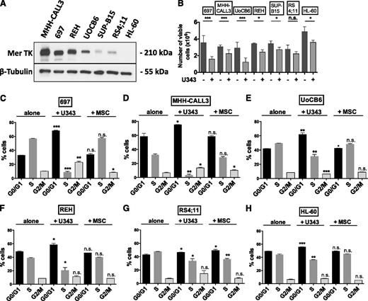 Fig. 2 U343 coculture induces quiescence in Mer-expressing ALL cell lines (MHH-CALL3, 697, REH, UoCB6, SUP-B15, and RS4;11). (Krause S, et al., 2015)
CD56 has a good positive predictive value for the diagnosis of myeloma and correlates well with bone lesions, and may be correlated with the pathogenesis of lytic lesions.
MHH-CALL-3 cells were established from the bone marrow of an 11-year-old girl diagnosed with pre B cell acute lymphoblastic leukemia (ALL) at the time of diagnosis in 1993, serving as a relevant model to study this specific subtype of leukemia.
By studying MHH-CALL-3 cells, researchers can gain insights into the disease progression, genetic aberrations, and cellular responses that underpin pre B cell ALL, aiding in the development of targeted therapeutic strategies.
The MHH-CALL-3 cells are utilized in preclinical studies, drug screening assays, and molecular investigations to further our understanding of pre B cell ALL progression and test potential therapeutic interventions.
Average Rating: 4.7 | 3 Scientist has reviewed this product
Helpful
Cells are the foundation of research, and Creative Bioarray's products and services have been a great help to me.
19 Oct 2022
Ease of use
After sales services
Value for money
Exceptional research tools
The MHH-CALL-3 cell products from Creative Bioarray have been instrumental in my research on pre B cell acute lymphoblastic leukemia (ALL).
10 Jan 2024
Ease of use
After sales services
Value for money
Valuable tools
These cells accurately model the disease phenotype and have facilitated in-depth molecular and genetic studies, providing valuable insights into disease progression. I highly recommend these cell products for researchers studying pre B cell ALL.
17 Oct 2023
Ease of use
After sales services
Value for money
Customer Support & Price Inquiry
