Featured Products
Our Promise to You
Guaranteed product quality, expert customer support

L-540
- Specification
- Technical Resources
- Recommended Products
- Publications
CSF1PO 11,13
D5S818 11,13
D7S820 11,12
D13S317 9
D16S539 11,13
TH01 7,9
TPOX 9,10
vWA 17,18
b. Cell-Based assay
c. In vivo efficacy study
Viruses: ELISA: reverse transcriptase negative; PCR: EBV +, HBV -, HCV -, HHV-8 -, HIV-1 -, HIV-2 -, HTLV-I/II -, MLV -, SMRV -
Store in liquid nitrogen.
Biosafety classification is based on U.S. Public Health Service Guidelines, it is the responsibility of the customer to ensure that their facilities comply with biosafety regulations for their own country.,
- Background
- Scientific Data
- Q & A
- Customer Review
- Product Information Sheet
The cultures L-540 were taken from the peripheral blood and bone marrow of a 20-year-old woman in the clinical stage IV B of Hodgkin's disease of the histologically proven nodular sclerosing type. The peripheral blood picture showed an eosinophilia of 25%, these specimens were taken in the prefinal stage of the disease. Establishing L-540 cells from the bone marrow of a patient with Hodgkin lymphoma allows researchers to investigate these cells' characteristics, behavior, and response under controlled laboratory conditions.
L-540 cells have been extensively characterized and have contributed to our understanding of the biology of Hodgkin lymphoma. They exhibit several features typical of Hodgkin and Reed-Sternberg (HRS) cells, the characteristic abnormal cells found in Hodgkin lymphoma. These features include large size, multi-lobulated nuclei, prominent nucleoli, and abundant cytoplasm. L-540 cells also express specific cell surface markers associated with Hodgkin lymphoma, such as CD30 and CD15.
L-540 cells have been used to study the cellular and molecular mechanisms underlying Hodgkin's lymphoma. Researchers have utilized these cells to investigate disease progression, oncogenic pathways, and the interactions between the tumor cells and the microenvironment. These cells have been employed in screening potential drugs and therapeutic agents for treating Hodgkin's lymphoma. They offer a valuable platform for testing drug efficacy, elucidating drug resistance mechanisms, and identifying potential novel treatment approaches.
Chromosome Instability in Hodgkin Lymphoma Cells Associated with Sister Chromatid Cohesion Defects
Aberrant sister chromatid cohesion causes chromosome instability (CIN) and thus contributes to the development of cancer. The study confirms that L-540 cell lines exhibit CIN phenotypes and are aneuploid. The L-540 cell lines were found to be aneuploid compared to the diploid control. The total chromosome numbers ranged from 41 to 148, with a model chromosome number of 56, and normal chromosomes ranging from 56 to 74.
Cohesion normally functions by tethering nascent synthesized chromatids together to prevent premature segregation and thus chromosome instability. Cohesion was categorized into normal (cohesed) or aberrant (primary constriction gap; PCG). PCG is a visually appreciable gap between the sister chromatids within the DAPI channel. To further characterize the severity of the PCG phenotypes, all spreads exhibiting aberrant cohesion were further classified into three distinct sub-categories ranging from mild (PCGI) to moderate (PCGII) to severe (PCGIII). In L-540, the cohesion was 44.6%, while PCGI, PCGII, and PCGIII were 14.9%, 10.6%, and 29.9%, respectively (Fig. 1).
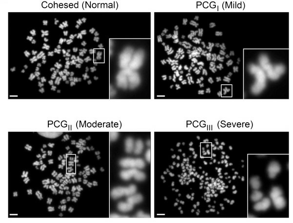 Fig. 1 Representative images of mitotic chromosome spreads from the L-540 cells. (Sajesh BV, et al., 2013)
Aberrant Expression of HLX in Hodgkin Lymphoma
NKL homeobox genes regulate cell and tissue differentiation. The HLX gene, encoded by NKL, exhibits abnormal activity in patients with Hodgkin's lymphoma (HL) cells. Here, genomic profiling (Fig. 2A) and fluorescence in situ hybridization (Fig. 2B) were performed to examine if HLX is abnormally activated in L-540.
To identify potential upstream factors of HLX, expression profiling of L-540 was conducted compared to HL cell lines. The results indicated activation of JAK-STAT signaling with high statistical significance. To examine directly the impact of STAT factors, the study quantified expression levels and performed siRNA-mediated knockdown of STAT5A, STAT5B, STAT3, and STAT4 (Fig. 3). STAT3 knockdown resulted in decreased HLX transcript levels, demonstrating that STAT3 activated the expression of HLX (Fig. 3C).
Overall, the study investigations excluded chromosomal rearrangements of the HLX locus at 1q41 (Fig. 2A) and demonstrated that STAT3 operated directly as a transcriptional activator of the HLX gene (Fig. 3). These analyses indicate that aberrantly expressed NKL homeobox gene HLX is part of a pathological gene network in HL, driving deregulated B-cell differentiation and survival.
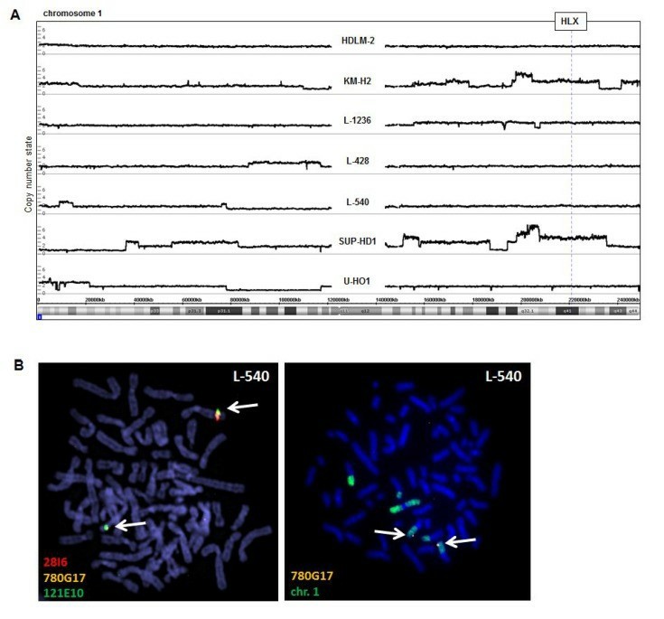 Fig. 2 (A) Genomic profiling was performed for seven HL cell lines, including L-540 cell lines. (B) Fluorescence in situ hybridization (FISH) in L-540 demonstrated wild-type configurations at the locus of HLX although chromosome 1 showed some structural abnormalities. (Nagel S, et al., 2018)
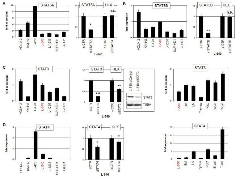 Fig. 3 RQ-PCR analysis of STAT-factors in HL cell lines, including L-540 cell lines. (Nagel S, et al., 2018)
Lestaurtinib Inhibiting Proliferation and Inducing Apoptosis in Hodgkin Lymphoma
JAK/STAT constitutive activation plays an important role in the pathogenesis of HL. Lestaurtinib is an orally bioavailable multi-kinase inhibitor recently shown to inhibit JAK2. The study analyzed the effect of Lestaurtinib treatment in L-540 cell lines.
Proliferation and apoptosis in response to Lestaurtinib of cultured HL cells were evaluated in L-540 cell lines after 48 h of treatment and compared to cells treated with DMSO vehicle control (normalized to 100%). At 300 nM of Lestaurtinib, a 66% reduction in proliferation was observed in L-540. At 300 nM of doxorubicin, the reduction in proliferation was 54% in L-540 (Fig. 4). At 300 nM, apoptosis increased by 10% in L-540. No significant differences were observed between 48, 72, or 96 hours.
The JAK/STAT pathway is one of the most frequently altered pathways in HL. To assess in greater detail the effects of Lestaurtinib-mediated JAK2 inhibition on the JAK2/STAT5 signaling pathway, protein levels of STAT5, phosphor-STAT5, STAT3, and phosphor-STAT3 were then analyzed. Following 1 hour of 300 nM of Lestaurtinib treatment, phosphor-STAT5, and phosphor-STAT3 levels decreased respectively (Fig. 5). After 1 h of 300 nM of Lestaurtinib treatment, Bcl-xL mRNA expression levels had decreased by 37% in L-540 (Fig. 6). This downregulation of Bcl-xL could explain the proapoptotic effect of Lestaurtinib.
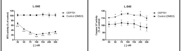 Fig. 4 Proliferation (left) and apoptosis (right) analysis after 48 h of Lestaurtinib treatment in L-540 cell lines. (Diaz T, et al., 2011)
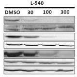 Fig. 5 Western blot analysis of JAK2/STAT5 pathway protein levels in L-540 cells after 1 h of Lestaurtinib treatment at different doses: 30, 100, and 300 nM. (Diaz T, et al., 2011)
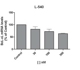 Fig. 6 Bcl-xL mRNA analysis after 1 h of Lestaurtinib treatment in L-540 cell lines. (Diaz T, et al., 2011)
L-540 cells are derived from the bone marrow of a 20-year-old woman with Hodgkin lymphoma.
L-540 cells are associated with the nodular sclerosis subtype of Hodgkin lymphoma.
Yes, L-540 cells are frequently used as a model for investigating drug sensitivity and testing potential therapies for Hodgkin lymphoma.
L-540 cells exhibit characteristics of Reed-Sternberg cells, the hallmark cells of Hodgkin lymphoma. They are known to express CD15 and CD30 antigens and produce cytokines, such as interleukin-6 (IL-6), that contribute to the pathogenesis of Hodgkin lymphoma.
Average Rating: 5.0 | 3 Scientist has reviewed this product
Satisfied
I recently purchased the L-540 cell line from Creative Bioarray to study the molecular mechanisms underlying Hodgkin lymphoma. Overall, I am quite satisfied with the product. The cells arrived promptly, and the packaging was secure. The provided documentation was helpful and detailed, guiding me through the protocols for culturing and maintaining the cells.
11 Jan 2024
Ease of use
After sales services
Value for money
Effective assistance
I experienced difficulties growing L-540 cells. I contacted Creative Bioarray's customer service for assistance, and they guided me to resolve the issue. I am satisfied with the outcome.
02 Jan 2024
Ease of use
After sales services
Value for money
Remarkable growth and stability
The provided instructions from Creative Bioarray were comprehensive and user-friendly, allowing us to easily establish and maintain the cell line in our laboratory. The L-540 cells showed remarkable growth and stability, which has been crucial for our experiments.
11 Jan 2023
Ease of use
After sales services
Value for money
Customer Support & Price Inquiry
