In Vitro Neurotoxicity
Creative Bioarray now provides in vitro neurotoxicity assays including neurite outgrowth assay and high throughput microelectrode array to study the effects of chemicals on the morphology and electrophysiological activity of neurons.
Neurotoxicity represents negative effects of chemicals on the nervous system that can lead to prolonged or even permanent troublesome consequences, especially on the developing central nervous system. These consequences can include headache, delusions, loss of memory and/or vision, seizures, behavioral changes, mental retardation, and other mental issues, which, when revealed, are usually irreversible after a long term exposure.
Due to the imperceptibility of neurotoxicity, most of the project closures due to neurotoxic issues occur in the clinical phase, while safety issues related to other organs can be addressed much earlier in the drug development process. Late stage project closure leads not only to higher cost of time and money for drug developers, but also to greater risks for patients. Therefore, evaluating toxic effects of drugs on the nervous systems is of great importance in toxicological studies.
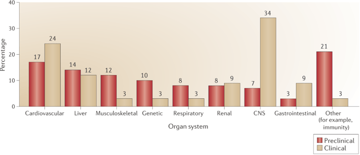 Figure 1. Project closures due to safety issues1.
Compared to animal tests (for example, TG 418, TG 419, and TG424), cell-based in vitro assays are way faster and much cheaper, which can also provide more information on the mechanisms at the same time, thus are more suitable for early stage high throughput screening.
Creative Bioarray provides in vitro neurotoxicity assays based on cultured neural cells for greater predictivity. We use automated high-content imaging or live-content imaging for neurite outgrowth assay to monitor early markers of neurotoxicity, and microelectrode array to accurately monitor the electrophisiological activity of the neurons.
- Neurite outgrowth assay
- Cytotoxicity assay
- Calcium flux assay
- High throughput multielectrode array
- Blood brain barrier (BBB) permeability
- ATP bioluminescence
Neurite (dendrite and axon) outgrowth is critical for communication between neurons, and key to neurogenesis and nervous system functioning, thus can serve as neurotoxic indicator. By incubating Neuronal cultures with test compounds and then staining the neuron microtubules via immunochemical assay, the length and number of neurites can be measured and counted using high-content imaging.
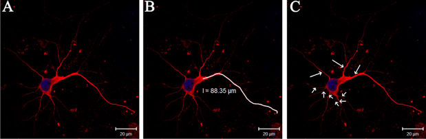 Figure 2. Detection of neurites by immunofluorescence2.
(A) Neurites (red) and nucleus (blue). (B) Manually tracing the length of the longest neurite (white) with the LSM (4.2.0.121) software. (C) Primary neurites (arrows) calculated.
Apart from neurite outgrowth assay, Creative Bioarray can also evaluate neurotoxicity by monitoring the effect of compounds on cell viability, apoptosis, membrane integrity, mitochondrial dysfunction, and oxidative stress.
Intracellular calcium flux has emergingly become an indicator for evaluating neurons response to pharmacological agents. Our service allows monitoring changes in intracellular calcium levels of neural cells in real-time.
High throughput Multielectrode Array (MEA) serves as a powerful technique to detect neurotoxic response. As neuron function is based on conducting electrical impulses via potentials, MEA assay can measure cytotoxicity and assess the effects of drugs both on neurite growth and their function.
Blood brain barrier (BBB) is the main route for most drug molecules entering in the central nervous system and it prevents many drugs from eliciting toxicity to the brain. Therefore, BBB permeability can be used to estimate the neurotoxicity of drugs to predict whether test compounds can reach the central nervous system in an amount sufficient to cause toxicity.
Creative Bioarray offers neurotoxicity kits, specifically designed to detect cytotoxicity in neural cells. This assay readout is via ATP bioluminescence.
With a group of highly experienced experts in the field, Creative Bioarray is capable of providing in vitro neurotoxicity assays of the highest quality. Toxicity assays on other tissues and organs are also available. If you have any special needs or questions regarding our services, please feel free to contact us to get support from our experts. We look forward to working with you in the near future.
References
- Cook, David, et al. "Lessons learned from the fate of AstraZeneca’s drug pipeline: a five-dimensional framework." Nat Rev Drug Discov 13.6 (2014): 419-431.
- Chen H, Lin W, Zhang Y, et al. IL-10 Promotes Neurite Outgrowth and Synapse Formation in Cultured Cortical Neurons after the Oxygen-Glucose Deprivation via JAK1/STAT3 Pathway[J]. Scientific Reports, 2016, 6.
